. 2010 Nov;106(5):675-86.
doi: 10.1093/aob/mcq169. Epub 2010 Sep 29.

Closure of plasmodesmata in maize (Zea mays) at low temperature: a new mechanism for inhibition of photosynthesis

Affiliations

Closure of plasmodesmata in maize (Zea mays) at low temperature: a new mechanism for inhibition of photosynthesis

Anna Bilska et al. Ann Bot. 2010 Nov.

Abstract

Background and aims: Photosynthesis is one of the processes most susceptible to low-temperature inhibition in maize, a tropical C4 crop not yet fully adapted to a temperate climate. C4 photosynthesis relies on symplasmic exchange of large amounts of photosynthetic intermediates between Kranz mesophyll (KMS) and bundle sheath (BS) cells. The aim of this study was to test the hypothesis that the slowing of maize photosynthesis at low temperature is related to ultrastructural changes in the plasmodesmata between KM and BS as well as BS and vascular parenchyma (VP) cells.

Methods: Chilling-tolerant (CT) KW 1074 and chilling-sensitive (CS) CM 109 maize (Zea mays) lines were studied. The effect of moderate chilling (14 °C) on the rate of photosynthesis, photosynthate transport kinetics, and the ultrastructure of plasmodesmata linking the KMS, BS and VP cells were analysed. Additionally, the accumulation of callose and calreticulin was studied by the immunogold method.

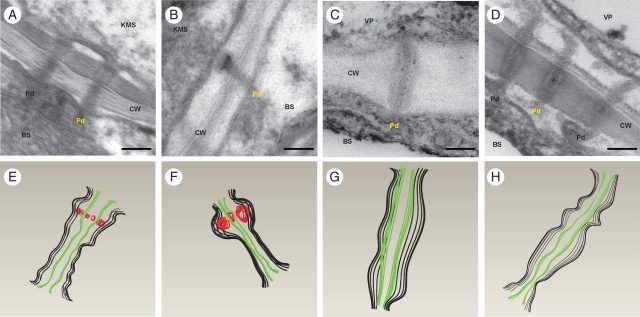
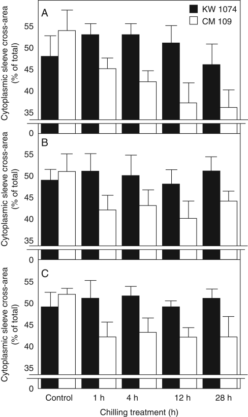
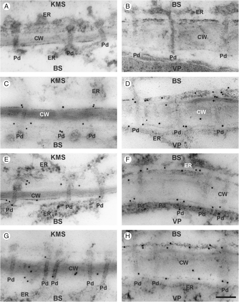
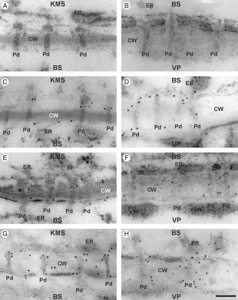
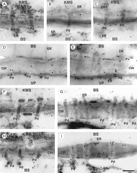
Key results: Chilling inhibited photosynthesis, photosynthate transfer to the phloem and photosynthate export from leaves in both lines. This inhibition was reversible upon cessation of chilling in the CT line but irreversible in the CS line. Simultaneously to physiological changes, chilling induced swelling of the sphincters of plasmodesmata linking KMS and BS cells and a decreased lumen of the cytoplasmic sleeve of plasmodesmata at the BS/VP interface in the CS line but not in the CT line. Accumulation of calreticulin, which occurred near the neck region of the closed plasmodesmata was observed after just 4 h of chilling and over-accumulation of callose at the KMS/BS and BS/VP interfaces occurred after 28 h of chilling.

Conclusions: Stronger chilling sensitivity of the CM 109 maize line compared with the KW 1074 line, shown by decreased photosynthesis and assimilate export from a leaf, is related to changes in the ultrastructure of leaf plasmodesmata at low temperature. The chain of reactions to chilling is likely to include calreticulin action resulting in rapid and efficient closure of the plasmodesmata at both KMS/BS and BS/VP interfaces. Callose deposition in a leaf was a secondary effect of chilling.


Figures

Fig. 1.
Fig. 1.
Cross-section of small bundle in a juvenile maize leaf. KMS, Kranz mesophyll; BS, bundle sheath; VP, vascular parenchyma; CC, companion cell; ○, thin-walled sieve tube; •, thick-walled sieve tube; X, xylem. Scale bar = 5 µm.
Fig. 2.
Fig. 2.
Longitudinal sections of plasmodesmata in control and chilled leaves of the CM 109 maize inbred line: (A–D) electron micrograms; (E–H) corresponding 3D shapes, designed with 3D CAD software, of the plasmodesmata shown in the micrograms in (A) to (D) (see Materials and methods), in which black lines = cytoplasmic sleeves, green lines = desmotubules and red lines = internal sphincters. (A, E) KMS/BS plasmodesmata in control leaves, showing an electron-dense ring consisting of regular elements (internal sphincter) at the KMS side. (B, F) KMS/BS cell plasmodesmata in chilled leaves of CM 109 line, showing a strongly enlarged internal sphincter. (C, G) BS/VP plasmodesmata in control leaves. (D, H) BS/VP plasmodesmata in chilled leaves – strong constriction, particularly at neck regions. KMS, Kranz mesophyll; BS, bundle sheath; VP, vascular parenchyma; CW, cell wall; ER, endoplasmic reticulum; Pd, plasmodesmata. Scale bar = 100 nm.
Fig. 3.
Fig. 3.
Cross-area of cytoplasmic sleeve of plasmodesmata in control and chilled (1 h, 4 h, 12 h, 28 h) leaves of two maize inbred lines: (A) plasmodesmata at bundle sheath–vascular parenchyma (BS/VP) interface; (B) plasmodesmata at bundle sheath–bundle sheath (BS/BS) interface; (C) plasmodesmata at vascular parenchyma–vascular parenchyma (VP/VP) interface. KW 1074, chilling-tolerant line and CM 109, chilling-sensitive line, as indicated. Values are percentages of whole cross-area of plasmodesmata, including plasmalemma. Values are means of 40–50 plasmodesmata ± s.e.
Fig. 4.
Fig. 4.
Immunogold localization of callose in control and chilled (4 h, 28 h) leaves of the KW 1074 maize inbred line (CT): (A, B) negative control – no immunogold labelling when primary antibody was omitted; (C, E, G) KMS/BS interface; (D, F, H) BS/VP interface. Note some callose labelling in the cell wall close to the plasmodesma in control leaves (C, D) and those chilled for 4 h (E, F) and 28 h (G, H). Abbreviations: CT, chilling-tolerant line; KMS, Kranz mesophyll; BS, bundle sheath; VP, vascular parenchyma; CW, cell wall; ER, endoplasmic reticulum; Pd, plasmodesmata. Scale bar = 100 nm.
Fig. 5.
Fig. 5.
Immunogold localization of callose in control and chilled (4 h, 28 h) leaves of the CM 109 maize inbred line (CS): (A, B) negative control; no immunogold labelling when primary antibody was omitted; (C, E, G) KMS/BS interface; (D, F and H) BS/VP interface. Note some callose labelling in cell wall close to the plasmodesma region in control leaves (C, D) and those chilled for 4 h (E, F). Note strong callose labelling near the plasmodesmata in leaves chilled for 28 h (G, H). Abbreviations: CS, chilling-sensitive line; KMS, Kranz mesophyll; BS, bundle sheath; VP, vascular parenchyma; CW, cell wall; ER, endoplasmic reticulum; Pd, plasmodesmata. Scale bar = 100 nm.
Fig. 6.
Fig. 6.
Immunogold localization of calreticulin in control and chilled (4 h) leaves of KW 1074 (CT) and CM 109 (CS) maize inbred lines: (A) negative control – no immunogold labelling when primary antibody was omitted; (B–E) CT line showing the KMS/BS (B, C) and BS/VP (D, E) interfaces in which calreticulin localizes mostly to ER and CW in both control (B, D) and chilled (C, E) leaves; (F–I) CS line showing KMS/BS (F) and BS/VP (G) interfaces in control leaves, in which calreticulin localizes mostly to ER and CW, and KMS/BS (H) and BS/VP (I) interfaces in chilled leaves, in which calreticulin accumulates at the neck regions of the plasmodesmata, mostly at the BS side. Abbreviations: CT, chilling-tolerant line; CS, chilling-sensitive line; KMS, Kranz mesophyll; BS, bundle sheath; VP, vascular parenchyma; CW, cell wall; ER, endoplasmic reticulum; Pd, plasmodesmata. Scale bar = 100 nm.

References

    1. Anisimov AV, Egorov AG. Plasmodesmata as a modulator of osmotic water fluxes in plants. Russian Journal of Plant Physiology. 2002;49:677–684.
    1. Aoki N, Hirose T, Takahashi S, Ono K, Ishimaru K, Ohsugi R. Molecular cloning and expression analysis of a gene for a sucrose transporter in maize (Zea mays L.) Plant and Cell Physiology. 1999;40:1072–1078. - PubMed
    1. Baluška F, Šamaj J, Napier R, Volkmann D. Maize calreticulin localizes preferentially to plasmodesmata in root apex. The Plant Journal. 1999;19:481–488. - PubMed
    1. Blackmann LM, Harper JDI, Overall RL. Localization of a centrin-like protein to higher plant plasmodesmata. European Journal of Cell Biology. 1999;78:297–304. - PubMed
    1. Botha CEJ, Cross RHM. Towards reconciliation of structure with function in plasmodesmata: who is gatekeeper? Micron. 2000;31:713–721. - PubMed