

Visual map development: bidirectional signaling, bifunctional guidance molecules, and competition
- PMID: 20880989
- PMCID: PMC2964178
- DOI: 10.1101/cshperspect.a001768
Visual map development: bidirectional signaling, bifunctional guidance molecules, and competition
Abstract
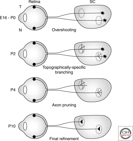
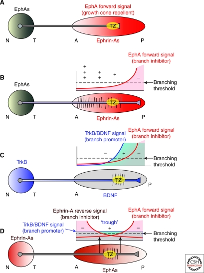
Topographic maps are a two-dimensional representation of one neural structure within another and serve as the main strategy to organize sensory information. The retina's projection via axons of retinal ganglion cells to midbrain visual centers, the optic tectum/superior colliculus, is the leading model to elucidate mechanisms of topographic map formation. Each axis of the retina is mapped independently using different mechanisms and sets of axon guidance molecules expressed in gradients to achieve the goal of representing a point in the retina onto a point within the target. An axon's termination along the temporal-nasal mapping axis is determined by opposing gradients of EphAs and ephrin-As that act through their forward and reverse signaling, respectively, within the projecting axons, each of which inhibits interstitial branching, cooperating with a branch-promoting activity, to generate topographic specific branching along the shaft of the parent axons that overshoot their correct termination zone along the anterior-posterior axis of the target. The dorsal-ventral termination position is then determined using a gradient of ephrin-B that can act as a repellent or attractant depending on the ephrin-B concentration relative to EphB levels on the interstitial branches to guide them along the medial-lateral axis of the target to their correct termination zone, where they arborize. In both cases, axon-axon competition results in axon mapping based on relative rather than absolute levels of repellent or attractant activity. The map is subsequently refined through large-scale pruning driven in large part by patterned retinal activity.
Figures


References
-
- Braisted JE, McLaughlin T, Wang HU, Friedman GC, Anderson DJ, O’Leary DDM 1997. Graded and lamina-specific distributions of ligands of EphB receptor tyrosine kinases in the developing retinotectal system. Dev Biol 191: 14–28 - PubMed
-
- Brown A, Yates PA, Burrols P, Ortuno D, Vaidya A, Jessell TM, Pfaff SL, O’Leary DDM, Lemke G 2000. Topographic mapping from the retina to the midbrain is controlled by relative but not absolute levels of EphA receptor signaling. Cell 102: 77–88 - PubMed
Publication types
MeSH terms
Substances
Grants and funding
LinkOut - more resources
Full Text Sources
Research Materials