

doi: 10.1128/JVI.01322-10.
Epub 2010 Sep 29.
Intrinsic cytoskeleton-dependent clustering of influenza virus M2 protein with hemagglutinin assessed by FLIM-FRET
Affiliations
- PMID: 20881046
- PMCID: PMC2976392
- DOI: 10.1128/JVI.01322-10
Item in Clipboard
Intrinsic cytoskeleton-dependent clustering of influenza virus M2 protein with hemagglutinin assessed by FLIM-FRET
J Virol.
2010 Dec.
Abstract
The hemagglutinin (HA) of influenza virus organizes the virus bud zone, a domain of the plasma membrane enriched in raft lipids. Using fluorescence lifetime imaging microscopy-fluorescence resonance energy transfer (FLIM-FRET), a technique that detects close colocalization of fluorescent proteins in transfected cells, we show that the viral proton channel M2 clusters with HA but not with a marker for inner leaflet rafts. The FRET signal between M2 and HA depends on the raft-targeting signals in HA and on an intact actin cytoskeleton. We conclude that M2 contains an intrinsic signal that targets the protein to the viral bud zone, which is organized by raft-associated HA and by cortical actin.
Figures
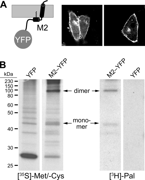
Generation and characterization of M2-YFP. (A) Scheme of M2-YFP in the membrane (gray) and expression of M2-YFP on the surface of transfected CHO cells. (B) Metabolic labeling of cells expressing YFP or M2-YFP with [35S]methionine/[35S]cysteine and [3H]palmitate for 6 h as indicated, immunoprecipitation with anti-green fluorescent protein (GFP) antibody, SDS-PAGE under nonreducing conditions, and fluorography. Positions of the M2-YFP monomer and dimer are marked with arrows. The M2 is from influenza A/Duck/Ukraine/1/63 (H3N8).
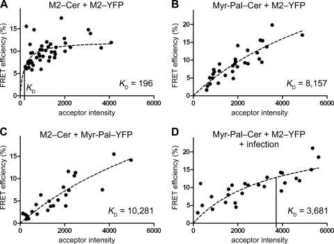
M2 associates with itself but not with membrane rafts. (A) Self-association of M2-Cer and M2-YFP evaluated by FLIM-FRET. The FRET efficiency E in the plasma membrane of each analyzed cell is plotted versus the acceptor intensity F; a hyperbolic function (equation 1, dashed line) is fitted to the data. The KD value to assess clustering is indicated as a vertical line on the x axis. Number of cells (n), 44; mean FRET efficiency ± standard error of the mean (SEM), 9.9% ± 0.4%; KD ± SEM, 196 ± 78. (B) FLIM-FRET of the raft marker Myr-Pal-Cer as the FRET donor with M2-YFP as the FRET acceptor. n, 31; E ± SEM, 9.0% ± 0.8%; KD ± SEM, 8,157 ± 4,679. (C) FLIM-FRET of M2-Cer as the FRET donor with the raft marker Myr-Pal-YFP as the FRET acceptor. n, 25; E ± SEM, 5.7% ± 0.8%; KD ± SEM, 10,281 ± 7,935. (D) FLIM-FRET of M2-YFP with Myr-Pal-Cer in virus-infected cells. Cells were transfected and 20 h later infected with fowl plague virus (H7N1) at a multiplicity of infection (MOI) of 40. FLIM-FRET measurements were performed 4 to 7 h postinfection. n, 26; E ± SEM, 10.2% ± 0.8%; KD ± SEM, 3,681 ± 1,964. Fluorescence was recorded with an Olympus FluoView 1000 confocal microscope, and FLIM was performed with the LSM upgrade kit and SymPhoTime software (PicoQuant).
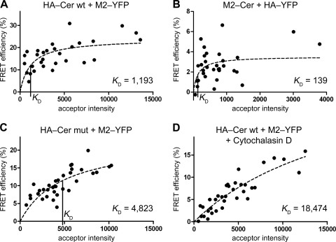
Clustering of M2 with HA. (A) FLIM-FRET analysis (as described in the legend to Fig. 2) with the wild-type HA-Cer (HA-Cer wt) as the FRET donor and M2-YFP as the FRET acceptor. n, 30; E ± SEM, 16.3% ± 1.2%; KD ± SEM, 1,193 ± 509. (B) FLIM-FRET analysis with M2-Cer as the FRET donor and HA-YFP as the FRET acceptor. n, 32; E ± SEM, 2.6% ± 0.3%; KD ± SEM, 139 ± 112. (C) FLIM-FRET of HA-Cer mut with M2-YFP. n, 33; E ± SEM, 10.6% ± 0.7%; KD ± SEM, 4,823 ± 1,882. (D) FLIM-FRET of HA-Cer wt with M2-YFP in the presence of cytochalasin D (1 μM, added 20 h prior to the measurements). n, 35; E ± SEM, 6.4% ± 0.7%; KD ± SEM, 18,474 ± 8,647. The KD value to assess clustering is indicated as a vertical line on the x axis where applicable. The HA of fowl plague virus (H7N1) was used in the analysis. Relative donor and acceptor intensity values did not differ significantly (P > 0.01, unpaired, two-sided Student's t test) between the experiments involving HA-Cer wt (panel A; 100%), HA-Cer mut (panel C; 73%), and HA-Cer wt plus cytochalasin D (panel D; 122%) as the FRET donor and M2-YFP as the acceptor (100, 106, and 100%, respectively), showing that the use of the HA mutant and cytochalasin D treatment did not significantly alter surface expression of the probes.
References
-
- Anderson, R. G., and K. Jacobson. 2002. A role for lipid shells in targeting proteins to caveolae, rafts, and other lipid domains. Science 296:1821-1825. - PubMed
Publication types
MeSH terms
Substances
LinkOut - more resources
Full Text Sources
