. 2010 Dec;21(23):4184-96.
doi: 10.1091/mbc.E10-02-0105. Epub 2010 Sep 29.

Nuclear organization and dynamics of 7SK RNA in regulating gene expression

Affiliations

Nuclear organization and dynamics of 7SK RNA in regulating gene expression

Kannanganattu V Prasanth et al. Mol Biol Cell. 2010 Dec.

Abstract

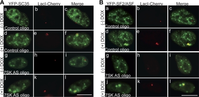
Noncoding RNAs play important roles in various aspects of gene regulation. We have identified 7SK RNA to be enriched in nuclear speckles or interchromatin granule clusters (IGCs), a subnuclear domain enriched in pre-mRNA processing factors. 7SK RNA, in association with HEXIM 1 and 2, is involved in the inhibition of transcriptional elongation by RNA polymerase II. Inhibition occurs via sequestration of the active P-TEFb kinase complex (CDK 9 and Cyclin T1/T2a/b or K) that is involved in phosphorylating the C-terminal domain of RNA polymerase II. Our results demonstrate that knock-down of 7SK RNA, by specific antisense oligonucleotides, results in the mislocalization of nuclear speckle constituents in a transcription-dependent manner, and the transcriptional up-regulation of a RNA polymerase II transcribed reporter gene locus. Furthermore, 7SK RNA transiently associates with a stably integrated reporter gene locus upon transcriptional down-regulation and its presence correlates with the efficient displacement of P-TEFb constituents from the locus. Our results suggest that 7SK RNA plays a role in modulating the available level of P-TEFb upon transcriptional down-regulation by sequestering its constituents in nuclear speckles.


Figures

Figure 1.
Figure 1.
7SK RNA is enriched in nuclear speckles. (Aa–Ad) Deconvolved images of RNA-FISH performed on HeLa cells using a 7SK oligonucleotide probe reveals enrichment of 7SK RNA (red, Aa) in nuclear speckles and colocalizes (Ac) with a known nuclear speckle marker, SF2/ASF (green, Ab). (Ba–Bd) Inhibition of RNA polymerase II by α-amanitin treatment shows 7SK RNA (red, Ba) enrichment in rounded up nuclear speckles (Bc). (Ca–Cc) Transient expression of an MS2 stem-loop tagged 7SK RNA (green, Ca) and MS2-BP-YFP in live HeLa cells shows colocalization of tagged 7SK RNA with SC35-RFP (red, Cb) in nuclear speckles (merge, Cc). DNA was stained with 4′,6-diamidino-2-phenylindole (DAPI) (blue, Ad, Bd). The scale bar represents 10 μm.
Figure 2.
Figure 2.
Knock-down of 7SK RNA results in the disorganization of nuclear speckle components. (A) Antisense oligonucleotides designed against 7SK RNA were used to knock-down 7SK in HeLa cells. 90% knock-down was achieved as assessed by Q-PCR. Control oligonucleotide had no effect. 7SK RNA levels were normalized to β-actin mRNA and are presented relative to RNA levels in scrambled oligonucleotide transfected cells. The data are shown as mean and SD values of three measurements per data point. (B) YFP-SF2/ASF (green, Ba) and 7SK RNA FISH (red, Bb) show prominent colocalization in speckles in HeLa cells treated with control antisense oligonucleotides (control oligo). Antisense knock-down of 7SK results in the appearance of large intranuclear foci of SF2/ASF (green, Bc, see arrowheads) and its disorganization from speckles. (C) YFP-SF2/ASF (green, Ca) and SC35 immunofluorescence (red, Cb) show prominent colocalization in speckles (merge, Cc) in HeLa cells treated with scrambled antisense oligonucleotides (control oligo). Antisense knock-down of 7SK results in the appearance of large intranuclear foci of SF2/ASF (green, Cd) and its disorganization from speckles. The cell that shows SF2/ASF foci also shows appearance of small rounded up SC35 foci (see arrows, red, Ce). Interestingly, both SF2/ASF and SC35 foci do not colocalize (merge, Cf). (D) YFP-SF2/ASF (green, Da) and immunolocalization of B” U2snRNP (red, Db) show prominent colocalization at the speckles in HeLa cells treated with control antisense oligonucleotides (control oligo). Cells lacking 7SK RNA demonstrate intranuclear foci of SF2/ASF (green, Dc). However, B” U2 snRNP shows a more homogenous distribution but continues to localize in Cajal bodies (red, Dd, see arrow). (E) RNA FISH using oligo dT probes shows poly(A+) RNA enrichment in speckles of control oligonucleotide treated cells (red, Eb). In contrast, following 7SK knock-down poly (A+) RNA appears as a more homogenous nuclear pool (red, Ed). Scale bar represents 5 μm.
Figure 3.
Figure 3.
Displacement of splicing factors from nuclear speckles upon 7SK depletion is transcription dependent. (A) Immunofluorescence localization of CDK9 (green, Aa) and the hyperphosphorylated form of RNA pol II (H14 ab, red, Ab) in control oligonucleotide treated U2OS cell shows a punctate nuclear localization excluding the nucleoli (arrow). 7SK antisense olgonucleotide-treated cell shows a reorganization of the distribution of CDK9 to nucleoli (green, Ac), whereas the RNA pol II localization remains unaltered (red, Ad). (B) Immunoblot analysis using antibodies against P-TEFb components and splicing factors in HeLa cells shows no change in their levels in control versus 7SK AS treated cells. (C) YFP-SF2/ASF (green, Ca) and B” (red, Cb) localization in control oligonucleotide treated HeLa cells followed by α-amanitin incubation shows that YFP-SF2/ASF is enriched around the nucleoli whereas B” protein localizes to the rounded up speckles (merge, Cc). α-amanitin incubated 7SK antisense oligonucleotide treated HeLa cells also shows a similar distribution of YFP-SF2/ASF and B” (Ce–Ch) indicating the disorganization of splicing factors from nuclear speckles observed in 7SK depleted cells is transcription dependent. DNA is counterstained with DAPI (blue, Cd, Ch). The bar indicates 10 μm.
Figure 4.
Figure 4.
7SK depletion up-regulates reporter gene expression using the pTet-ON system. (A) Schematic representation of the reporter construct used for the analysis. The arrows in the figure indicate the positions of the primer used for the RT-PCR analysis. The figure is adapted and modified from (Janicki et al., 2004). (B) In control cells MS2-YFP localization shows an accumulation of the reporter RNA at the site of transcription 15 min post gene activation (green, Bc, arrowhead). In the absence of 7SK RNA, higher levels of reporter RNA accumulate at the transcription site (green, Bd, arrow). Scale bar represents 5 μm. (C) RT-PCR analysis of RNA samples from cells isolated 15 min and 30 min post DOX treatment shows up-regulation of the reporter gene in cells that are depleted of 7SK RNA (D) Immunoblot analysis using an anti-GFP antibody demonstrates the up-regulation of reporter CFP protein in 7SK knock-down cells corroborating the increased transcriptional activation of the reporter gene in the absence of 7SK RNA. α-tubulin was used as a loading control.
Figure 5.
Figure 5.
The recruitment of SR-family splicing factors to a transcriptionally active locus remains unaffected in cells that are depleted of 7SK RNA. In U2OS 2-6-3-TOLC cells, YFP-SC35 (A) and YFP-SF2/ASF (B) are recruited to the transcriptionally active locus ([+] DOX, Ad–f; Aj–l; Bd–f; Bj–l) in the presence (YFP-SC35, Ad–f; YFP-SF2/ASF, Bd–f) or absence (YFP-SC35, Aj–l; YFP-SF2/ASF, Bj–l) of 7SK RNA. Note that the 7SK depleted cells show prominent round YFP-SC35 (Ag–l) and YFP-SF2/ASF (Bg–l) subnuclear foci. The scale bar represents 10 μm.
Figure 6.
Figure 6.
P-TEFb, but not 7SK, is present at sites of active transcription of a stably integrated reporter locus. (A and B) Immunolocalization to CDK9 (red, Aa) and 7SK RNA-FISH (red, Ba) in U2OS 2-6-3 tet-ON cells. These constituents (Ac, Bc) are not visualized at the inactive locus (LacI-YFP, green, Ab and Bb). (C and D) 240 min after Dox addition and induction of transcription CDK9 (red, Ca), but not 7SK RNA (red, Da), is concentrated at the active locus (green, Cb and Db). While CDK9 (Cc) shows direct overlap with decondensed chromatin, 7SK RNA (Dc) is absent from the region. (E and F) CDK9 (red, Ea) but not 7SK RNA (red, Fa) is enriched at the actively transcribing reporter gene locus (green, Eb and Fb) as observed by MS2-YFP bound to nascent RNA (Ec and Fc). DNA is counterstained with DAPI (blue, Ed, Fd). Scale bar represents 10 μm.
Figure 7.
Figure 7.
7SK transiently accumulates at the reporter gene locus during cessation of transcription. Immunolocalization of CDK9 (green; Ab) and 7SK RNA FISH (red; Ac) in representative U2OS 2-6-3 tet-OFF cells demonstrates that at the marked locus (LacI-CFP, blue; Aa) CDK9, but not 7SK RNA, is present during active transcription (Aa–Ad). At 2 min 30s post Dox addition to turn the locus “OFF” CDK9 (Bb) is still detectable at the locus (Ba) and a slight enrichment in 7SK RNA (Bc) that colocalizes with CDK9 (Bd) is now apparent. Five minutes post-Dox perfusion 7SK RNA (Cc) is now highly enriched at the locus (Ca) and colocalizes strongly with CDK9 (Cd). At 7 min 30 s post-Dox perfusion, the marked chromatin locus (Da) shows decrease in both CDK9 (Db) and 7SK RNA (Dc) signals, with only partial colocalization (Dd). By 10 min post-Dox perfusion neither CDK9 (Eb) nor 7SK RNA (Ec) is present at the marked locus (Ea). Scale bar represents 5 μm.
Figure 8.
Figure 8.
7SK RNA depletion affects the displacement of Cdk9 from the reporter gene locus during transcriptional silencing. Immunolocalization of CDK9 (Ab; Aa–Ja) in U2OS 2-6-3 tet-OFF cells demonstrates that in the majority of control oligo treated cells (A–E), CDK9 is displaced within 10–15 min post DOX addition (Ca–c). However, in cells that are depleted of 7SK RNA (F–J) CDK9 continues to associate with the gene locus for a longer duration of time post-DOX addition (H–I). DNA is counterstained with DAPI (Ac–Jc). The bar represents 5 μm. (K) Histogram showing reduced displacement of CDK9 from the gene locus in cells that are transfected with ASO against 7SK RNA (n = 50). Scale bar represents 5 μm.

References

    1. Amaral P. P., Dinger M. E., Mercer T. R., Mattick J. S. The eukaryotic genome as an RNA machine. Science. 2008;319:1787–1789. - PubMed
    1. Barboric M., Kohoutek J., Price J. P., Blazek D., Price D. H., Peterlin B. M. Interplay between 7SK snRNA and oppositely charged regions in HEXIM1 direct the inhibition of P-TEFb. EMBO J. 2005;24:4291–4303. - PMC - PubMed
    1. Barboric M., Lenasi T., Chen H., Johansen E. B., Guo S., Peterlin B. M. 7SK snRNP/P-TEFb couples transcription elongation with alternative splicing and is essential for vertebrate development. Proc. Natl. Acad. Sci. USA. 2009;106:7798–7803. - PMC - PubMed
    1. Barrandon C., Bonnet F., Nguyen V. T., Labas V., Bensaude O. The transcription-dependent dissociation of P-TEFb-HEXIM1-7SK RNA relies upon formation of hnRNP-7SK RNA complexes. Mol. Cell Biol. 2007;27:6996–7006. - PMC - PubMed
    1. Barrandon C., Spiluttini B., Bensaude O. Non-coding RNAs regulating the transcriptional machinery. Biol. Cell. 2008;100:83–95. - PubMed

Publication types

MeSH terms

LinkOut - more resources