Comparative Study
. 2010 Sep 29;30(39):12950-7.
doi: 10.1523/JNEUROSCI.1158-10.2010.

Membrane estrogen receptors stimulate intracellular calcium release and progesterone synthesis in hypothalamic astrocytes

Affiliations
Comparative Study

Membrane estrogen receptors stimulate intracellular calcium release and progesterone synthesis in hypothalamic astrocytes

John Kuo et al. J Neurosci. .

Abstract

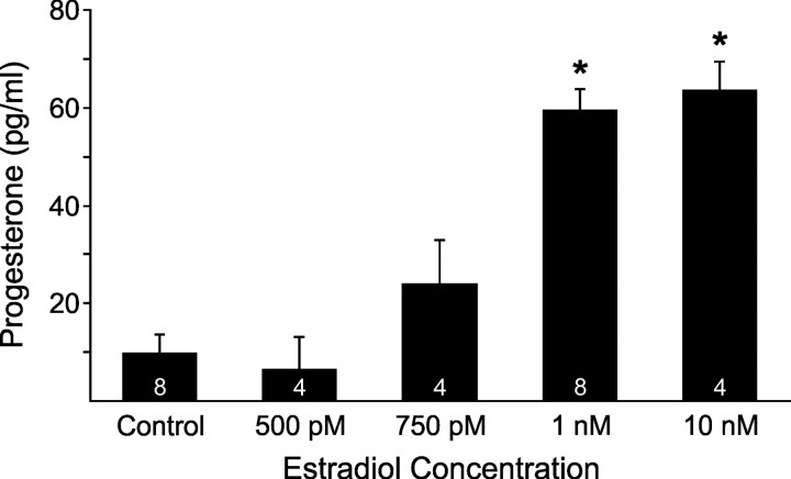
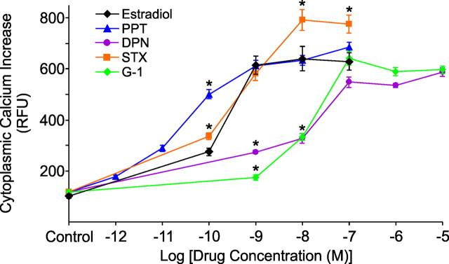
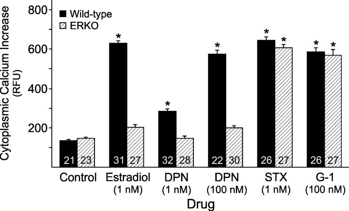
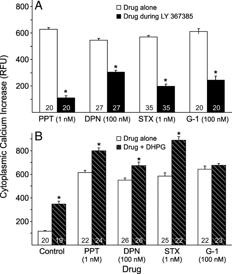
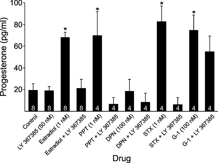
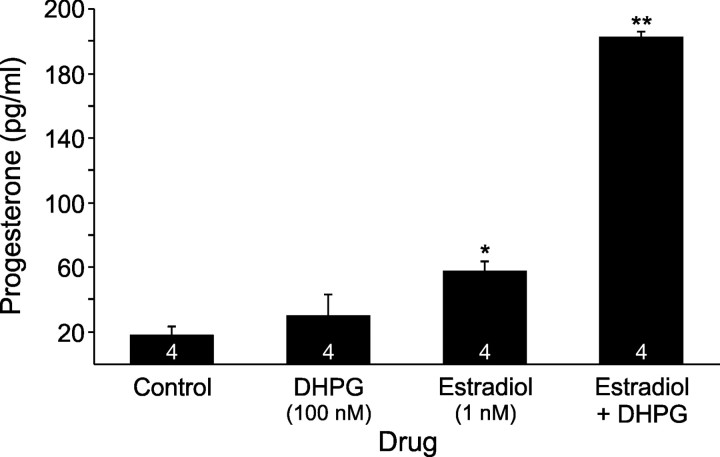
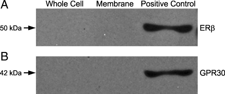
In hypothalamic astrocytes obtained from adult female rats, estradiol rapidly increased free cytoplasmic calcium concentrations ([Ca(2+)](i)) that facilitate progesterone synthesis. The present study demonstrated that estradiol (1 nm) significantly and maximally stimulated progesterone synthesis within 5 min, supporting a rapid, nongenomic mechanism. The group I metabotropic glutamate receptor (mGluR1a) antagonist LY 367385 [(S)-(+)-a-amino-4-carboxy-2-methylbenzeneacetic acid] attenuated both the estradiol-induced [Ca(2+)](i) release and progesterone synthesis. To investigate membrane-associated estrogen receptors (mERs), agonists for ERα, ERβ, STX-activated protein, and GPR30 were compared. The selective ERα agonist propylpyrazole triole (PPT) and STX most closely mimicked the estradiol-induced [Ca(2+)](i) responses, where PPT was more potent but less efficacious than STX. Only high doses (100 nm) of selective ERβ agonist diarylpropionitrile (DPN) and GPR30 agonist G-1 induced estradiol-like [Ca(2+)](i) responses. With the exception of DPN (even at 100 nm), all agonists stimulated progesterone synthesis. The PPT- and STX-induced [Ca(2+)](i) release and progesterone synthesis were blocked by LY 367385. While the G-1-stimulated [Ca(2+)](i) release was blocked by LY 367385, progesterone synthesis was not. Since GPR30 was detected intracellularly but not in the membrane, we interpreted these results to suggest that G-1 could activate mGluR1a on the membrane and GPR30 on the smooth endoplasmic reticulum to release intracellular calcium. Although STX and G-1 maximally stimulated [Ca(2+)](i) release in astrocytes from estrogen receptor-α knock-out (ERKO) mice, estradiol in vivo did not stimulate progesterone synthesis in the ERKO mice. Together, these results indicate that mERα is mainly responsible for the rapid, membrane-initiated estradiol-signaling that leads to progesterone synthesis in hypothalamic astrocytes.


Figures

Figure 1.
Figure 1.
Dose–response relationship between estradiol stimulation and progesterone synthesis in adult hypothalamic astrocytes. Estradiol at 1 nm, 10 nm, 100 nm (62.6 ± 7.0 pg/ml, n = 4), and 1 μm (64.6 ± 6.5 pg/ml, n = 4) all stimulated significant and maximal progesterone synthesis (p < 0.05 vs control) that were similar to each other (p > 0.05). However, estradiol at 100 pm (10.4 ± 6.0 pg/ml, n = 4), 250 pm (10.1 ± 10.1 pg/ml, n = 4), 500 pm, and 750 pm failed to stimulate progesterone synthesis (p > 0.05 vs control). The number of samples for each estradiol concentration is indicated in the bar graph. Significantly different compared with control (*p < 0.05, one-way ANOVA with Student–Newman–Keuls post hoc test).
Figure 2.
Figure 2.
Comparison of dose–response relationships measured in [Ca2+]i between estradiol and several ER agonists in adult hypothalamic astrocytes. At 100 pm, the [Ca2+]i response for PPT and STX were both greater than estradiol (p < 0.05). At 1 nm, PPT and STX exposure resulted in an estradiol-like [Ca2+]i release (p > 0.05), whereas DPN and G-1 responses were significantly lower than estradiol (p < 0.05). At 10 nm, PPT again induced [Ca2+]i levels similar to estradiol (p > 0.05), STX had a greater [Ca2+]i response compared with estradiol (p < 0.05), and DPN and G-1 continued to have a weaker [Ca2+]i response (p < 0.05). At 100 nm, PPT, DPN, and G-1 all induced an estradiol-like [Ca2+]i release (p > 0.05), while STX continued to induce a greater [Ca2+]i response (p < 0.05). There was no difference between unstimulated controls (p > 0.05). For all drug concentrations, a range of 14–33 astrocytes were imaged. Significantly different compared with estradiol at the same concentration (*p < 0.05, one-way ANOVA with Student–Newman–Keuls post hoc test).
Figure 3.
Figure 3.
Importance of mERα for the estradiol-induced [Ca2+]i release in adult hypothalamic astrocytes. The estradiol (1 nm)-induced [Ca2+]i release was attenuated in the ERKO mouse astrocytes (p > 0.05 vs control). DPN (1 and 100 nm) did not induce a [Ca2+]i response (p > 0.05 vs control) in ERKO astrocytes. Wild-type and ERKO astrocytes were able to respond normally and equally to STX at 1 nm and G-1 at 100 nm (p < 0.05 vs control). The number of astrocytes for each drug concentration is indicated in the bar graph. Significantly different compared with control (*p < 0.05, one-way ANOVA with Student–Newman–Keuls post hoc test).
Figure 4.
Figure 4.
Effect of the mGluR1a on ER agonist-induced [Ca2+]i responses in adult hypothalamic astrocytes. In A, the [Ca2+]i levels induced with PPT (1 nm), DPN (100 nm), STX (1 nm), and G-1 (100 nm) were all significantly blocked by LY 367385 (20 nm; p < 0.05). In B, concurrent DHPG (100 nm) administration significantly enhanced the [Ca2+]i levels induced by PPT (1 nm; p < 0.05), DPN (100 nm; p < 0.05), and STX (1 nm; p < 0.05). However, the [Ca2+]i response induced with G-1 (100 nm) was not enhanced with DHPG (100 nm) exposure (p > 0.05). The number of astrocytes for each drug concentration is indicated in the bar graph. Significantly different when comparing drugs (control, PPT, DPN, STX, and/or G-1) in the absence or presence of LY 367385 in A (*p < 0.05, paired Student's t test) or in the absence or presence of DHPG in B (*p < 0.05, unpaired Student's t test).
Figure 5.
Figure 5.
Effect of LY 367385, mGluR1a antagonist, on progesterone synthesis upon exposure to estradiol and several mER agonists in adult hypothalamic astrocytes. LY 367385 (50 nm) by itself did not influence progesterone synthesis with levels similar to control (p > 0.05). Estradiol (1 nm), PPT (1 nm), STX (1 nm), and G-1 (100 nm) stimulated significantly greater progesterone synthesis than control (p < 0.05), but DPN (100 nm) did not (p > 0.05 vs control). Progesterone produced by exposure to estradiol (1 nm), PPT (1 nm), and STX (1 nm) was blocked by LY 367385 (50 nm; p < 0.05). However, the G-1 (100 nm)-stimulated progesterone synthesis was not attenuated by LY 367385 (50 nm; p > 0.05). The number of samples for each drug concentration is indicated in the bar graph. Significantly different compared with control (*p < 0.05, one-way ANOVA with Student–Newman–Keuls post hoc test).
Figure 6.
Figure 6.
Effect of DHPG, mGluR1a agonist, on estradiol stimulated progesterone synthesis in adult hypothalamic astrocytes. Estradiol at 1 nm stimulated significant progesterone synthesis (p < 0.05 vs control or DHPG), but DHPG at 100 nm did not (p > 0.05 vs control). Estradiol (1 nm) in combination with DHPG (100 nm) stimulated a much higher progesterone level than estradiol (1 nm) or DHPG (100 nm) alone (p < 0.05). The number of samples for each drug concentration is indicated in the bar graph. Significantly different compared with control (*p < 0.05, one-way ANOVA with Student–Newman–Keuls post hoc test) or compared with all other groups (**p < 0.05, one-way ANOVA with Student–Newman–Keuls post hoc test).
Figure 7.
Figure 7.
Coimmunoprecipitation of ERβ-mGluR1a and GPR30-mGluR1a from cultured adult hypothalamic astrocytes. The lanes are representative of whole-cell lysate, plasma membrane fraction, and whole-cell lysate without coimmunoprecipitation as a positive control, respectively. In A, ERβ-mGluR1a coimmunoprecipitation with mGluR1a antibodies used to pull-down the receptor complex and antibodies against ERβ used for detection. In B, GPR30-mGluR1a coimmunoprecipitation with mGluR1a antibodies used in the pull-down phase and the complex detected with GPR30 antibodies. The mGluR1a did not coimmunoprecipitate with ERβ or GPR30 within the plasma membrane fraction or whole-cell lysate.

References

    1. Acconcia F, Ascenzi P, Bocedi A, Spisni E, Tomasi V, Trentalance A, Visca P, Marino M. Palmitoylation-dependent estrogen receptor alpha membrane localization: regulation by 17beta-estradiol. Mol Biol Cell. 2005;16:231–237. - PMC - PubMed
    1. Beyer C, Raab H. Nongenomic effects of oestrogen: embryonic mouse midbrain neurones respond with a rapid release of calcium from intracellular stores. Eur J Neurosci. 1998;10:255–262. - PubMed
    1. Bologa CG, Revankar CM, Young SM, Edwards BS, Arterburn JB, Kiselyov AS, Parker MA, Tkachenko SE, Savchuck NP, Sklar LA, Oprea TI, Prossnitz ER. Virtual and biomolecular screening converge on a selective agonist for GPR30. Nat Chem Biol. 2006;2:207–212. - PubMed
    1. Bondar G, Kuo J, Hamid N, Micevych P. Estradiol-induced estrogen receptor-alpha trafficking. J Neurosci. 2009;29:15323–15330. - PMC - PubMed
    1. Boulware MI, Weick JP, Becklund BR, Kuo SP, Groth RD, Mermelstein PG. Estradiol activates group I and II metabotropic glutamate receptor signaling, leading to opposing influences on cAMP response element-binding protein. J Neurosci. 2005;25:5066–5078. - PMC - PubMed

Publication types

MeSH terms

LinkOut - more resources