. 2010 Dec;104(6):3293-304.
doi: 10.1152/jn.00663.2010. Epub 2010 Sep 29.

High ratio of synaptic excitation to synaptic inhibition in hilar ectopic granule cells of pilocarpine-treated rats

Affiliations

High ratio of synaptic excitation to synaptic inhibition in hilar ectopic granule cells of pilocarpine-treated rats

Ren-Zhi Zhan et al. J Neurophysiol. 2010 Dec.

Abstract

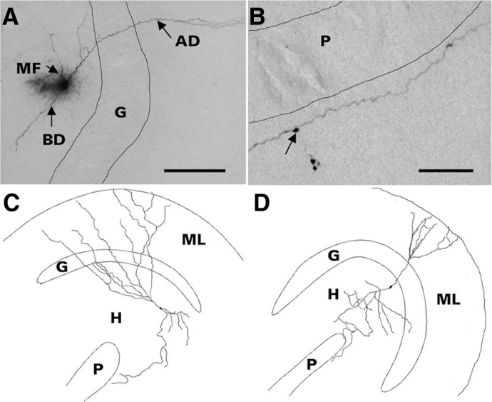
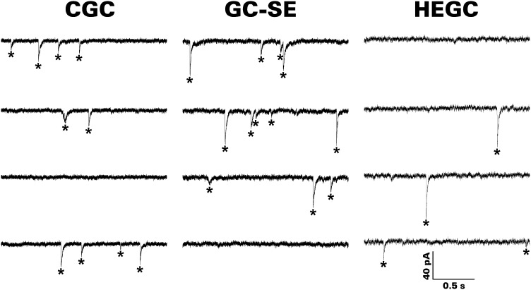
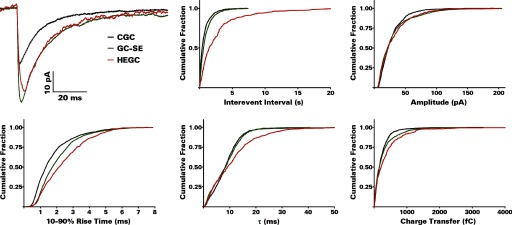
After experimental status epilepticus, many dentate granule cells born into the postseizure environment migrate aberrantly into the dentate hilus. Hilar ectopic granule cells (HEGCs) have also been found in persons with epilepsy. These cells exhibit a high rate of spontaneous activity, which may enhance seizure propagation. Electron microscopic studies indicated that HEGCs receive more recurrent mossy fiber innervation than normotopic granule cells in the same animals but receive much less inhibitory innervation. This study used hippocampal slices prepared from rats that had experienced pilocarpine-induced status epilepticus to test the hypothesis that an imbalance of synaptic excitation and inhibition contributes to the hyperexcitability of HEGCs. Mossy fiber stimulation evoked a much smaller GABA(A) receptor-mediated inhibitory postsynaptic currents (IPSC) in HEGCs than in normotopic granule cells from either control rats or rats that had experienced status epilepticus. However, recurrent mossy fiber-evoked excitatory postsynaptic currents (EPSCs) of similar size were recorded from HEGCs and normotopic granule cells in status epilepticus-experienced rats. HEGCs exhibited the highest frequency of miniature excitatory postsynaptic currents (mEPSCs) and the lowest frequency of miniature inhibitory postsynaptic currents (mIPSCs) of any granule cell group. On average, both mEPSCs and mIPSCs were of higher amplitude, transferred more charge per event, and exhibited slower kinetics in HEGCs than in granule cells from control rats. Charge transfer per unit time in HEGCs was greater for mEPSCs and much less for mIPSCs than in the normotopic granule cell groups. A high ratio of excitatory to inhibitory synaptic function probably accounts, in part, for the hyperexcitability of HEGCs.


Figures

Fig. 1.
Fig. 1.
Morphology of representative hilar ectopic granule cells (HEGCs). Cells were filled with biocytin during whole cell patch-clamp recording, fixed slices were cut into serial 60-μm-thick sections, biocytin was visualized with nickel-intensified avidin-horseradish peroxidase-diaminobenzidine (Vectastain Elite ABC kit, Vector Laboratories, Burlingame, CA), and cell morphology was reconstructed with use of Neurolucida (MicroBrightField, Williston, VT). Criteria for confirmation of HEGC identity included hilar location of the soma, apical dendrite(s) penetrating into or directed toward the dentate molecular layer, and axon with giant boutons in area CA3 and extensive branches within the hilus. A: the apical dendrite (AD) of this HEGC crosses the granule cell body layer (G) and penetrates the molecular layer. The mossy fiber (MF) branches within the hilus. This cell also has a basal dendrite (BD) directed into the hilus. Scale bar, 500 μm. B: HEGC mossy fiber courses through stratum lucidum of area CA3 adjacent to the pyramidal cell body layer (P). Arrow indicates a giant bouton from which a filopodium originates. Scale bar, 200 μm. C: reconstructed morphology of an HEGC from which miniature inhibitory postsynaptic currents (mIPSCs) were recorded. D: reconstructed morphology of an HEGC from which miniature excitatory postsynaptic currents (mEPSCs) were recorded. The apical dendrites of both cells reached the outer edge of the dentate molecular layer (ML) and the cell in D had a short basal dendrite directed into the hilus (H). The main branch of the mossy fiber reached stratum lucidum of area CA3.
Fig. 2.
Fig. 2.
Mossy fiber stimulation evokes a small GABAA receptor–mediated IPSC in HEGCs and a large GABAA receptor–mediated IPSC in GC-SEs. Top: responses recorded from a representative normotopic dentate granule cell from a control rat (CGC), a normotopic granule cell from a rat subjected to pilocarpine-induced status epilepticus (GC-SE), and an HEGC. The mossy fiber-evoked IPSC, NMDA receptor–mediated EPSC, and AMPA/kainate (KA) receptor–mediated EPSC were recorded sequentially. *Stimulus artifact. Bottom left: the mossy fiber-evoked IPSC was smallest in HEGCs whether measured by amplitude or charge transfer. IPSC charge transfer was greatest in GC-SEs largely because of the longer response duration. *P < 0.05 compared with CGC; **P < 0.001 compared with GC-SE and P = 0.025 compared with CGC by Newman-Keuls test after 1-way ANOVA yielded P < 0.001. ***P < 0.01 compared with CGC or GC-SE by Newman-Keuls test after 1-way ANOVA yielded P = 0.003. Bottom right: the charge transfer ratio of AMPA/kainate receptor–mediated EPSC (AMPA) to GABAA receptor–mediated IPSC (GABA) was significantly greater in HEGCs than in GC-SEs. However, the charge transfer ratios of NMDA receptor–mediated EPSC (NMDA) to AMPA/kainate receptor–mediated EPSC were not significantly different. *P = 0.01 by 2-tailed t-test.
Fig. 3.
Fig. 3.
mEPSCs are recorded more frequently from HEGCs than from either GC-SEs or CGCs; mEPSC amplitude is generally greater in HEGCs and GC-SEs than in CGCs. The traces shown are from representative experiments. Recordings were made with a CsCl-based internal solution, and the membrane potential was clamped at −70 mV. Individual events that met our criterion are indicated by asterisks.
Fig. 4.
Fig. 4.
Cumulative probability plots show lower interevent intervals (higher frequency) of mEPSCs in HEGCs and differences from control in mEPSC amplitude, rise time, decay time constant, and charge transfer per event. Results were computed from the number of events given in Table 1, which also presents the grouped data. Averaged mEPSCs recorded from a representative CGC, GC-SE, and HEGC are shown at the top left.
Fig. 5.
Fig. 5.
Histograms of 10–90% rise times for all mEPSCs and mIPSCs recorded in this study. The numbers of events included are given in Tables 1 and 2. In HEGCs, slowly rising mEPSCs (>4 ms) constituted a significantly higher percentage of total mEPSCs than in the normotopic granule cell groups, and there was no between-group difference in the percentage of rapidly rising mEPSCs (<1 ms). Slowly rising mIPSCs constituted a significantly higher percentage of total mIPSCs than in the normotopic granule cell groups, but rapidly rising mIPSCs constituted a significantly smaller percentage of the total in both HEGCs and GC-SEs than in CGCs.
Fig. 6.
Fig. 6.
mIPSCs are much less frequently recorded from HEGCs than from either GC-SEs or CGCs; mIPSC amplitude is generally greater in HEGCs and GC-SEs than in CGCs. The traces shown are from representative experiments. Recordings were made with a CsCl-based internal solution, and the membrane potential was clamped at −70 mV. Individual events that met our criterion are indicated by asterisks. In the 2nd segment of the CGC trace, the 1st asterisk marks 2 overlapping mIPSCs.
Fig. 7.
Fig. 7.
Cumulative probability plots show greater intervent intervals (lower frequency) of mIPSCs in HEGCs and differences from control in mIPSC amplitude, rise time, decay time constant, and charge transfer per event. Results were computed from the number of events given in Table 2, which also presents the grouped data. Averaged mIPSCs recorded from a representative CGC, GC-SE, and HEGC are shown at the top left.
Fig. 8.
Fig. 8.
Analysis of miniature synaptic events shows an excess of excitation over inhibition in HEGCs compared with the normotopic granule cell groups. mEPSCs and mIPSCs were recorded from different cells. The numbers of cells from which each were recorded are given in Tables 1 and 2. Event frequency and charge transfer/s were computed for each granule cell studied, and the results obtained from all cells in each group were averaged. The mean mEPSC frequency or charge transfer/s was divided by the mean mIPSC frequency or charge transfer/s. The mEPSC/mIPSC ratio for both measures was much greater in HEGCs.

Similar articles

Cited by

References

    1. Andre V, Marescaux C, Nehlig A, Fritschy JM. Alterations of hippocampal GABAergic system contribute to spontaneous recurrent seizures in the rat lithium pilocarpine model of temporal lobe epilepsy. Hippocampus 11: 452–468, 2001 - PubMed
    1. Bonde S, Ekdahl CT, Lindvall O. Long-term neuronal replacement in adult rat hippocampus after status epilepticus despite chronic inflammation. Eur J Neurosci 23: 965–974, 2006 - PubMed
    1. Brooks-Kayal AR, Shumate MD, Jin H, Rikhter TY, Coulter DA. Selective changes in single cell GABAA receptor subunit expression and function in temporal lobe epilepsy. Nat Med 4: 1166–1172, 1998 - PubMed
    1. Buckmaster PS, Dudek FE. Network properties of the dentate gyrus in epileptic rats with hilar neuron loss and granule cell axon reorganization. J Neurophysiol 77: 2685–2696, 1997 - PubMed
    1. Buckmaster PS, Jongen-Rêlo AL. Highly specific neuron loss preserves lateral inhibitory circuits in the dentate gyrus of kainate-induced epileptic rats. J Neurosci 19: 9519–9529, 1999 - PMC - PubMed

Publication types

MeSH terms

LinkOut - more resources