

IOP-induced lamina cribrosa displacement and scleral canal expansion: an analysis of factor interactions using parameterized eye-specific models
- PMID: 20881292
- PMCID: PMC3101679
- DOI: 10.1167/iovs.10-5500
IOP-induced lamina cribrosa displacement and scleral canal expansion: an analysis of factor interactions using parameterized eye-specific models
Abstract
Purpose: To study the anterior-posterior lamina cribrosa deformation (LCD) and the scleral canal expansion (SCE) produced by an increase in IOP and identify the main factors and interactions that determine these responses in the monkey.
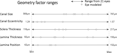
Methods: Eye-specific baseline models of the LC and sclera of both eyes of three normal monkeys were constructed. Morphing techniques were used to generate 888 models with controlled variations in LC thickness, position and modulus (stiffness), scleral thickness and modulus, and scleral canal size and eccentricity. Finite element modeling was used to simulate an increase in IOP from 10 to 15 mm Hg. A two-level, full-factorial experimental design was used to select factor combinations and to determine the sensitivity of LCD and SCE to the eight factors, independently and in interaction.
Results: LCD was between 53.6 μm (posteriorly) and -12.9 μm (anteriorly), whereas SCE was between 0.5 and 15.2 μm (all expansions). LCD was most sensitive to laminar modulus and position (24% and 21% of the variance in LCD, respectively), whereas SCE was most sensitive to scleral modulus and thickness (46% and 36% of the variance in SCE, respectively). There were also strong interactions between factors (35% and 7% of the variance in LCD and SCE, respectively).
Conclusions: IOP-related LCD and SCE result from a complex combination of factors, including geometry and material properties of the LC and sclera. This work lays the foundation for interpreting the range of individual sensitivities to IOP and illustrates that predicting individual ONH response to IOP will require the measurement of multiple factors.
Figures


References
-
- Burgoyne CF, Downs JC, Bellezza AJ, Suh JK, Hart RT. The optic nerve head as a biomechanical structure: a new paradigm for understanding the role of IOP-related stress and strain in the pathophysiology of glaucomatous optic nerve head damage. Prog Retin Eye Res. 2005;24:39–73 - PubMed
-
- Quigley HA. Glaucoma: macrocosm to microcosm the Friedenwald lecture. Invest Ophthalmol Vis Sci. 2005;46:2662–2670 - PubMed
-
- Sigal IA, Ethier CR. Biomechanics of the optic nerve head. Exp Eye Res. 2009;88:799–807 - PubMed
-
- Kirwan RP, Crean JK, Fenerty CH, Clark AF, O'Brien CJ. Effect of cyclical mechanical stretch and exogenous transforming growth factor-beta1 on matrix metalloproteinase-2 activity in lamina cribrosa cells from the human optic nerve head. J Glaucoma. 2004;13:327. - PubMed
-
- Sigal IA, Flanagan JG, Ethier CR. Factors influencing optic nerve head biomechanics. Invest Ophthalmol Vis Sci. 2005;46:4189–4199 - PubMed
Publication types
MeSH terms
Grants and funding
LinkOut - more resources
Full Text Sources
Other Literature Sources
