

TGFB1-induced extracellular expression of TGFBIp and inhibition of TGFBIp expression by RNA interference in a human corneal epithelial cell line
- PMID: 20881301
- PMCID: PMC3053104
- DOI: 10.1167/iovs.10-5362
TGFB1-induced extracellular expression of TGFBIp and inhibition of TGFBIp expression by RNA interference in a human corneal epithelial cell line
Abstract
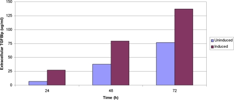
Purpose: To report the increased production of extracellular transforming growth factor β-induced protein (TGFBIp) by human corneal epithelial cells (HCECs) after induction by TGFB1 and the inhibition of TGFBIp production in induced and noninduced HCECs by RNA interference (RNAi).
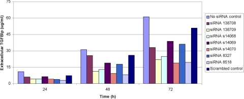
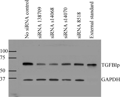
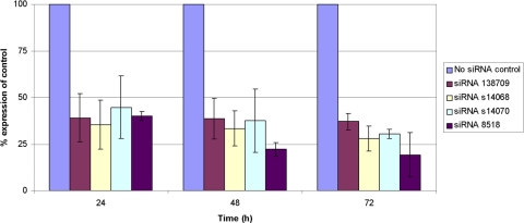
Methods: HCECs were cultured in serum-free medium and treated with 0 or 10 ng/mL TGFB1 over a period of 72 hours. Commercially available siRNAs targeting TGFBI mRNA were mixed with a transfection reagent and used to reverse transfect TGFB1-induced and noninduced HCECs. Extracellular and intracellular concentrations of TGFBIp were measured by ELISA and Western blot analysis, respectively, and TGFBI RNA was assayed using semiquantitative RT-PCR.
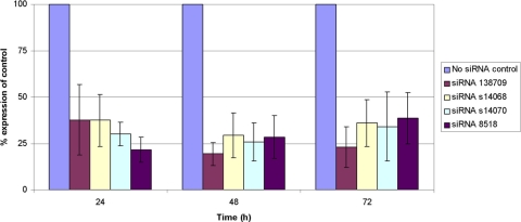
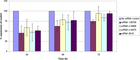
Results: HCECs constitutively express TGFBIp, and treatment with TGFB1 results in up to a fourfold increase in the amount of extracellular TGFBIp. Four commercially available siRNAs targeting TGFBI mRNA produced a >70% decrease in extracellular TGFBIp within 48 hours after transfection of noninduced HCECs but a <25% decrease in extracellular TGFBIp by 48 hours after transfection of TGFB1-induced HCECs. The suppression of extracellular TGFBIp production correlated with a decrease in intracellular TGFBIp production and TGFBI mRNA expression after transfection.
Conclusions: Extracellular TGFBIp expression by HCECs is increased several fold after exposure to TGFB1. Both HCEC-constitutive and HCEC-induced TGFBIp production can be inhibited with RNA interference, though the effect was greater and lasted longer for constitutive than induced TGFBIp production. Given that the corneal deposits in the TGFBI dystrophies consist of TGFBIp derived from HCECs, RNAi represents a potential means to inhibit primary dystrophic deposit formation and recurrence after surgical intervention.
Figures
Similar articles
-
Inhibition of TGFBIp expression by lithium: implications for TGFBI-linked corneal dystrophy therapy.Invest Ophthalmol Vis Sci. 2011 May 17;52(6):3293-300. doi: 10.1167/iovs.10-6405. Invest Ophthalmol Vis Sci. 2011. PMID: 21310903
-
Development of allele-specific gene-silencing siRNAs for TGFBI Arg124Cys in lattice corneal dystrophy type I.Invest Ophthalmol Vis Sci. 2014 Feb 18;55(2):977-85. doi: 10.1167/iovs.13-13279. Invest Ophthalmol Vis Sci. 2014. PMID: 24425855
-
Lysophosphatidic acid activates TGFBIp expression in human corneal fibroblasts through a TGF-β1-dependent pathway.Cell Signal. 2012 Jun;24(6):1241-50. doi: 10.1016/j.cellsig.2012.02.009. Epub 2012 Feb 21. Cell Signal. 2012. PMID: 22374302
-
Biochemical mechanisms of aggregation in TGFBI-linked corneal dystrophies.Prog Retin Eye Res. 2020 Jul;77:100843. doi: 10.1016/j.preteyeres.2020.100843. Epub 2020 Jan 29. Prog Retin Eye Res. 2020. PMID: 32004730 Review.
-
Clinical and genetic aspects of the TGFBI-associated corneal dystrophies.Ocul Surf. 2014 Oct;12(4):234-51. doi: 10.1016/j.jtos.2013.12.002. Epub 2014 Jul 18. Ocul Surf. 2014. PMID: 25284770 Review.
Cited by
-
Decoding cellular plasticity and niche regulation of limbal stem cells during corneal wound healing.Stem Cell Res Ther. 2024 Jul 6;15(1):201. doi: 10.1186/s13287-024-03816-y. Stem Cell Res Ther. 2024. PMID: 38971839 Free PMC article.
-
Transcriptome Analysis of TGFBI Knockdown vs Normal Corneal Epithelial Cells: Implications for TGFBI Corneal Dystrophy Treatment.Biochem Genet. 2025 Jul 15. doi: 10.1007/s10528-025-11191-3. Online ahead of print. Biochem Genet. 2025. PMID: 40665122
-
Characterization of a pluripotent stem cell-derived matrix with powerful osteoregenerative capabilities.Nat Commun. 2020 Jun 15;11(1):3025. doi: 10.1038/s41467-020-16646-2. Nat Commun. 2020. PMID: 32541821 Free PMC article.
-
Histone methylation levels correlate with TGFBIp and extracellular matrix gene expression in normal and granular corneal dystrophy type 2 corneal fibroblasts.BMC Med Genomics. 2015 Nov 9;8:74. doi: 10.1186/s12920-015-0151-8. BMC Med Genomics. 2015. PMID: 26553048 Free PMC article.
-
Association of TCF4 and CLU polymorphisms with Fuchs' endothelial dystrophy and implication of CLU and TGFBI proteins in the disease process.Eur J Hum Genet. 2012 Jun;20(6):632-8. doi: 10.1038/ejhg.2011.248. Epub 2012 Jan 11. Eur J Hum Genet. 2012. PMID: 22234156 Free PMC article.
References
-
- Brady SE, Rapuano CJ, Arentsen JJ, Cohen EJ, Laibson PR. Clinical indications for and procedures associated with penetrating keratoplasty. 1983–1988. Am J Ophthalmol. 1989;108(2):118–122 - PubMed
-
- Cosar CB, Sridhar MS, Cohen EJ, et al. Indications for penetrating keratoplasty and associated procedures: 1996–2000. Cornea. 2002;21(2):148–151 - PubMed
-
- Dobbins KR, Price FW, Jr, Whitson WE. Trends in the indications for penetrating keratoplasty in the midwestern United States. Cornea. 2000. ;19(6):813–816 - PubMed
-
- Lois N, Kowal VO, Cohen EJ, et al. Indications for penetrating keratoplasty and associated procedures: 1989–1995. Cornea. 1997;16(6):623–629 - PubMed
-
- Aldave AJ, Sonmez B. Elucidating the molecular genetic basis of the corneal dystrophies: are we there yet? Arch Ophthalmol. 2007;125(2):177–186 - PubMed
Publication types
MeSH terms
Substances
Grants and funding
LinkOut - more resources
Full Text Sources
Miscellaneous
