. 2010 Nov 4;468(7320):98-102.
doi: 10.1038/nature09387. Epub 2010 Sep 29.

Osteoclast differentiation factor RANKL controls development of progestin-driven mammary cancer

Affiliations

Osteoclast differentiation factor RANKL controls development of progestin-driven mammary cancer

Daniel Schramek et al. Nature. .

Abstract

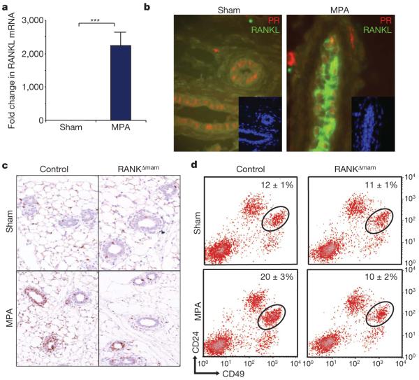
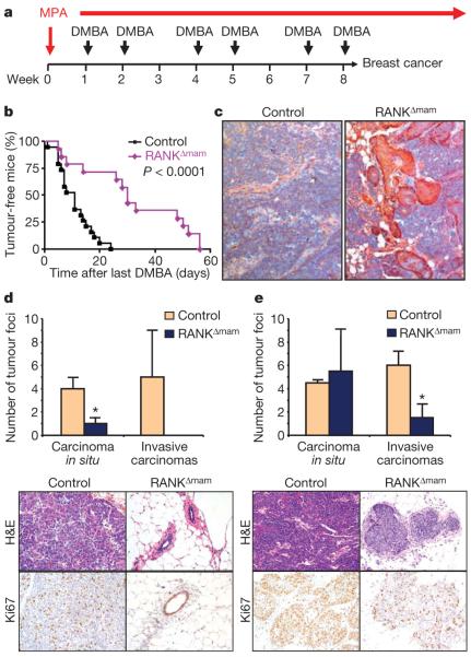
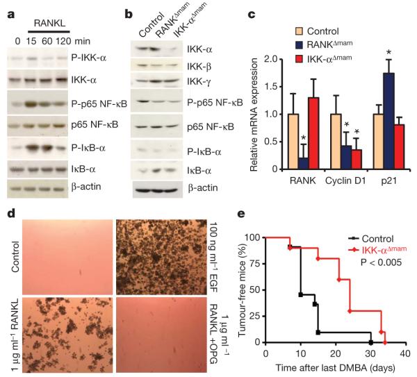
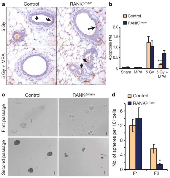
Breast cancer is one of the most common cancers in humans and will on average affect up to one in eight women in their lifetime in the United States and Europe. The Women's Health Initiative and the Million Women Study have shown that hormone replacement therapy is associated with an increased risk of incident and fatal breast cancer. In particular, synthetic progesterone derivatives (progestins) such as medroxyprogesterone acetate (MPA), used in millions of women for hormone replacement therapy and contraceptives, markedly increase the risk of developing breast cancer. Here we show that the in vivo administration of MPA triggers massive induction of the key osteoclast differentiation factor RANKL (receptor activator of NF-κB ligand) in mammary-gland epithelial cells. Genetic inactivation of the RANKL receptor RANK in mammary-gland epithelial cells prevents MPA-induced epithelial proliferation, impairs expansion of the CD49f(hi) stem-cell-enriched population, and sensitizes these cells to DNA-damage-induced cell death. Deletion of RANK from the mammary epithelium results in a markedly decreased incidence and delayed onset of MPA-driven mammary cancer. These data show that the RANKL/RANK system controls the incidence and onset of progestin-driven breast cancer.


Figures

Figure 1
Figure 1. The progesterone derivative MPA triggers in vivo RANKL expression and the proliferation of mammary epithelial cells through RANK
a, b, Induction of RANKL expression by the progesterone derivative MPA. Nulliparous wild-type females were implanted subcutaneously with slow-release MPA pellets or treated with sham surgery. a, mRNA encoding RANKL was determined in purified mammary epithelial cells by quantitative RT–PCR three days after implantation. β-Actin mRNA was used for normalization. Data are shown as fold change compared with sham treatment (means ± s.e.m., n = 3). b, In situ immunostaining of progesterone receptor (PR, red) and RANKL (green) in mammary epithelial cells after treatment with MPA for 3 days. c, Epithelial proliferation in mammary glands of control littermates and RANKΔmam females 3 days after sham treatment and MPA implantation. Proliferation was determined by in situ Ki67 immunostaining. d, Marked increase in the stem-cell-enriched CD24+CD49fhi population (MaSC) in MPA-treated mammary glands in control but not in RANKΔmam mammary glands. Representative FACS profiles showing CD24 and CD49f expression of lineage-negative (CD31 (endothelial cells) CD45 (haematopoietic cells) TER199 (erythroid cells)) mammary MaSCs from MPA-treated or sham-treated eight-week-old virgin females. Asterisk, P < 0.05; three asterisks, P < 0.001 (Student’s t-test).
Figure 2
Figure 2. RANK controls the incidence and onset of progestin-driven mammary cancer
a, Carcinogenesis scheme involving MPA and DMBA. Nulliparous six-week-old female mice were implanted subcutaneously with MPA pellets and treated orally with DMBA as indicated for eight weeks. b, Onset of palpable mammary tumours in MMTV-Cre rankfloxed females (RANKΔmam) (n = 14) and age-matched littermate control females (n = 19) treated with MPA pellets and DMBA as indicated in a. Data are shown as the percentage of tumour-free mice after the last DMBA challenge. Median tumour onset for controls was 11 days after the last DMBA treatment; for RANKΔmam females it was 30 days. c, Representative histological sections of mammary tumours isolated from control littermate and RANKΔmam females 22 days after the last DMBA treatment. Cytokeratin 5 staining is shown. Original magnifications ×20. d, e, Numbers of carcinomas in situ and invasive mammary cancers in control and RANKΔmam females on day 7 (d) and day 22 (e) after the final DMBA treatment. Data are shown as means±s.e.m. per mouse (n = 3 mice per genotype). All ten mammary glands were analysed for each mouse. Asterisk, P < 0.05 (Student’s t-test). Bottom panels show representative histological sections with typical invasive adenocarcinomas in the control females. For RANKΔmam females, normal acinar morphology (day 7) and a carcinoma in situ (day 22) are shown. Haematoxylin/eosin (H&E)-stained sections and immunostaining for the proliferation marker Ki67 are shown. Original magnifications ×20.
Figure 3
Figure 3. RANK induces NF-κB signalling and anchorage-independent growth
a, Western blotting for phosphorylated (P) IKK-α, total IKK-α, phosphorylated p65 NF-κB, total p65 NF-κB, phosphorylated IκB-α, and total IκB-α in isolated MECs in response to stimulation with RANKL (1 μg ml−1). β-Actin is shown as loading control. b, Western blotting for IKK-α, IKK-β, IKK-γ, phosphorylated p65 NF-κB, total p65 NF-κB, phosphorylated IκB-α and total IκB-α in pooled late-stage mammary adenocarcinomas (n = 4 for each lane) that developed in control, RANKΔmam and IKK-αΔmam females. β-Actin is shown as loading control. c, Expression of mRNA encoding RANK, cyclin D1 and p21 in late-stage mammary adenocarcinomas that developed in control, RANKΔmam and IKK-αΔmam females. Expression was determined by quantitative RT–PCR. β-Actin mRNA was used for normalization. Data are means±s.e.m. (n = 4 per group). d, Soft-agar colony formation assay. Growth of human SKBR3 breast cancer cells in soft agar in response to stimulation with RANKL (1 μg ml−1) or epidermal growth factor (100 ng ml−1). Anchorage-independent macroscopic colonies formed after 18 days in culture with RANKL, which was prevented by the decoy receptor osteoprogesterin (OPG) (1 μg ml−1). Controls were unstimulated SKBR3 cells. e, Onset of palpable mammary tumours in IKK-αΔmam (n = 10) and age-matched littermate control (n = 11) females treated with MPA pellets and DMBA. Data are shown as percentages of tumour-free mice after the last DMBA challenge. Median tumour onset for controls was 10 days after the last DMBA treatment, and for IKK-αΔmam females it was 24 days.
Figure 4
Figure 4. RANK protects from radiation-induced epithelial apoptosis and controls mammosphere formation
a, γ-Irradiation (5 Gy) induced mammary epithelial cell apoptosis in control and RANKΔmam female littermates either sham operated or implanted with a pellet of MPA. Apoptosis was detected by immunostaining for active caspase-3. Apoptotic nuclei of epithelial cells (arrows) are shown for representative mammary-gland sections. Original magnifications ×40. b, Quantification of mammary epithelial apoptosis. A minimum of 5,000 nuclei were counted for each mouse. Results shown are means±s.e.m. (n = 3 mice per group). Two asterisks, P < 0.02 (Student’s t-test). c, Self-renewal of mammary cancer stem cells (TICs) requires RANK expression. Mammary tumour cells from RANKΔmam and control littermate females treated with MPA and DMBA were cultured for 7 days; a small percentage of primary cells formed mammospheres (first passage). Primary mammospheres were then digested into single cells and assayed for their ability to form secondary mammospheres (second passage). Tumour cells from control littermate but not from RANKΔmam females could form secondary mammospheres determined 7 days after replating. d, Quantification of primary and secondary passage (F1 and F2) mammospheres from RANKΔmam and control littermate females treated with MPA and DMBA. Results shown are means±s.e.m. (n = 3 mice per group). Asterisk, P < 0.05 (Student’s t-test).

Comment in

References

    1. Jemal A, et al. Cancer statistics, 2009. CA Cancer J. Clin. 2009;59:225–249. - PubMed
    1. Rossouw JE, et al. Risks and benefits of estrogen plus progestin in healthy postmenopausal women: principal results from the Women’s Health Initiative randomized controlled trial. J. Am. Med. Assoc. 2002;288:321–333. - PubMed
    1. Beral V. Breast cancer and hormone-replacement therapy in the Million Women Study. Lancet. 2003;362:419–427. - PubMed
    1. Kong YY, et al. OPGL is a key regulator of osteoclastogenesis, lymphocyte development and lymph-node organogenesis. Nature. 1999;397:315–323. - PubMed
    1. Dougall WC, et al. RANK is essential for osteoclast and lymph node development. Genes Dev. 1999;13:2412–2424. - PMC - PubMed

Publication types

MeSH terms