. 2009 Sep 4;21(32-33):3419-24.
doi: 10.1002/adma.200801957.

The elusive path to cartilage regeneration

The elusive path to cartilage regeneration

Ernst B Hunziker. Adv Mater. .

Abstract

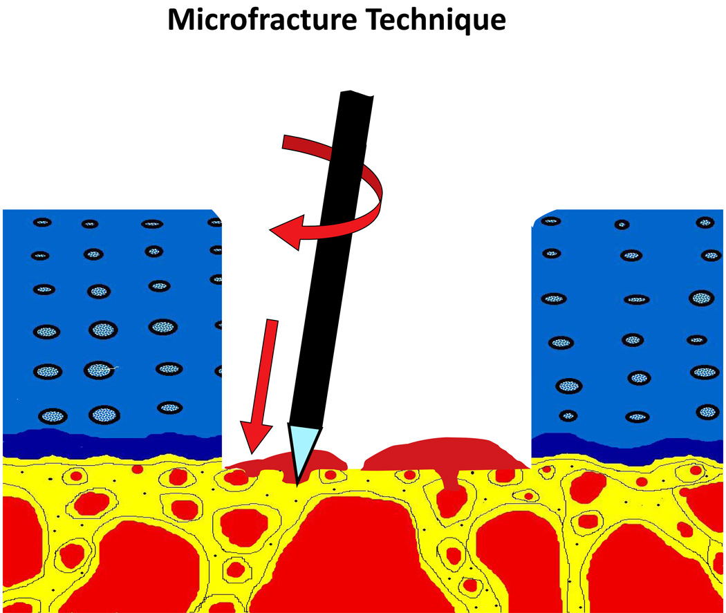
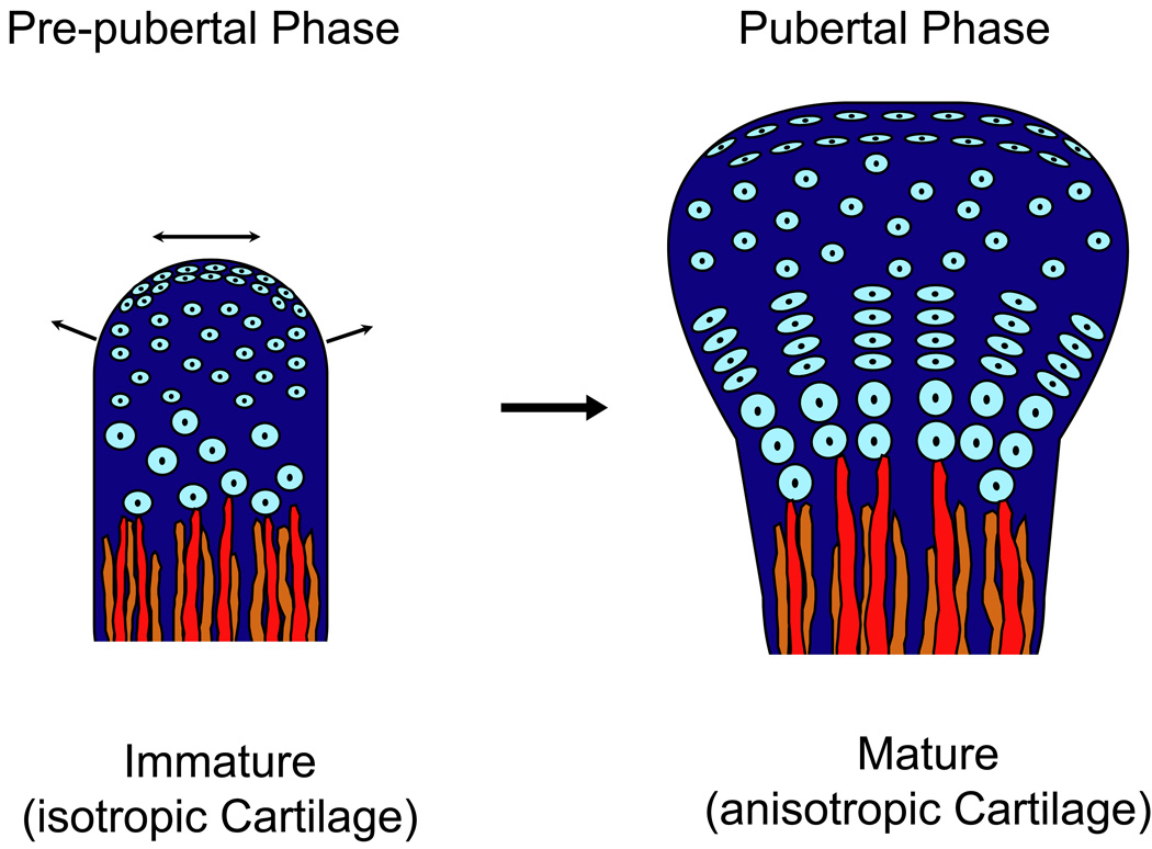
Numerous attempts have been made to develop an efficacious strategy for the repair of articular cartilage. These endeavours have been undaunted, if not spurred, by the challenge of the task and by the largely disappointing outcomes in animal models. Of the strategies that have been lately applied in a clinical setting, the autologous-chondrocyte-transplantation technique is the most notorious example. This methodology, which was prematurely launched on the clinical scene, was greeted with enthusiasm and has been widely adopted. However, a recent prospective and randomized clinical trial has revealed the approach to confer no advantage over conventional microfracturing. Why is the repair of articular cartilage such a seemingly intractable problem? The root of the evil undoubtedly lies in the tissue's poor intrinsic healing capacity. But the failure of investigators to tackle the biological stumbling blocks systematically rather than empirically is hardly a less inauspicious circumstance. Moreover, it is a common misbelief that the formation of hyaline cartilage per se suffices, whereas to be durable and functionally competent, the tissue must be fully mature. An appreciation of this necessity, coupled with a thorough understanding of the postnatal development of articular cartilage, would help to steer investigators clear of biological cul-de-sacs.


Figures

Fig. 1
Fig. 1
Schematic representation of the microfracturing technique. The floor of a shallow, full-thickness articular cartilage defect, viz., one that does not violate the subchondral bone plate, is perforated in several places using either a sharp instrument or a small-bore drill. By this action, the bone-marrow space is opened. Blood bearing mesenchymal stem cells percolates into the defect wherein it forms a haematoma.
Fig. 2
Fig. 2
Schematic representation of the ACI-technique. A fragment of articular cartilage is removed from the periphery of the joint that bears the lesion. This fragment of tissue is digested to release the chondrocytes, which are expanded in vitro for 11–21 days. A periosteal flap is then removed from the medial tibia and sutured over the lesion. A suspension of the expanded chondrocytes is injected beneath the flap into the lesion. Reproduced with the publisher’s permission from Brittberg et al. [32].
Fig. 3
Fig. 3
Light micrographs of vertical sections through the tibial condylar articular cartilage of rabbits aged 1 month (A), 2 months (B), 3 months (C) and 8 months (D). One month after birth (A), the articular cartilage is of an immature, foetal type, which is characterized by an isotropic organization of the chondrocytes. This immature type of tissue persists during the pre-pubertal growth spurt [up to 2 months (B)]. By the time the rabbit has attained sexual maturity [3 months (C)], the articular cartilage manifests the anisotropic structure that is so characteristic of the adult animal (D). During the course of postnatal development, the mature type of tissue does not evolve from the immature one by a process of gradual remodelling. The immature tissue is almost completely resorbed; only the superficial zone of chondroprogenitor cells is spared, and these cells give rise directly to mature chondrocytes that produce an adult type of articular cartilage. Reproduced with the publisher’s permission from Hunziker et al. [39].
Fig. 4
Fig. 4
Schematic representation of the sequence of events that lead to the formation of the cartilage anlage that develops into two synovial joints and three bones (A), and that are involved in its early postnatal development (B, C). A: Mesenchymal stem cells condense and differentiate into chondroblasts, which form an anlage of cartilage. In the interzone (boxed area), a transverse split develops, which is destined to become the synovial cavity. Chondroblasts within the interzone give rise to chondrocytes – which form a layer of immature articular cartilage – to fibrochondrocytes – which form the meniscus – and to synovial cells – which elaborate the synovium. B: During early postnatal development, the immature articular cartilage acts as a superficial growth plate for the radial expansion of the epyphyseal bone. Chondroprogenitor cells within the superficial zone proliferate exuberantly and differentiate into immature chondrocytes that produce an immature (isotropic) type of cartilage. During puberty, chondroprogenitor cells within the superficial zone continue to proliferate, but they are reprogrammed to differentiate into mature chondrocytes that produce an adult (anisotropic) type of cartilage. C: During puberty, the expanding core of epiphyseal bone (E) divides the cartilage tissue into a superficial articular layer and a growth plate proper. The latter then serves for the elongation and expansion of the metaphysis (M) and the diaphysis (not shown).
Fig. 4
Fig. 4
Schematic representation of the sequence of events that lead to the formation of the cartilage anlage that develops into two synovial joints and three bones (A), and that are involved in its early postnatal development (B, C). A: Mesenchymal stem cells condense and differentiate into chondroblasts, which form an anlage of cartilage. In the interzone (boxed area), a transverse split develops, which is destined to become the synovial cavity. Chondroblasts within the interzone give rise to chondrocytes – which form a layer of immature articular cartilage – to fibrochondrocytes – which form the meniscus – and to synovial cells – which elaborate the synovium. B: During early postnatal development, the immature articular cartilage acts as a superficial growth plate for the radial expansion of the epyphyseal bone. Chondroprogenitor cells within the superficial zone proliferate exuberantly and differentiate into immature chondrocytes that produce an immature (isotropic) type of cartilage. During puberty, chondroprogenitor cells within the superficial zone continue to proliferate, but they are reprogrammed to differentiate into mature chondrocytes that produce an adult (anisotropic) type of cartilage. C: During puberty, the expanding core of epiphyseal bone (E) divides the cartilage tissue into a superficial articular layer and a growth plate proper. The latter then serves for the elongation and expansion of the metaphysis (M) and the diaphysis (not shown).

Similar articles

Cited by

References

    1. Aigner T, Soder S. Z Rheumatol. 2008;67:32. - PubMed
    1. Richter W. Curr Opin Rheumatol. 2007;19:451. - PubMed
    1. Gelse K, Muhle C, Franke O, Park J, Jehle M, Durst K, Goken M, Hennig F, von der Mark K, Schneider H. Arthritis Rheum. 2008;58:475. - PubMed
    1. Park J, Gelse K, Frank S, von der Mark K, Aigner T, Schneider H. J Gene Med. 2006;8:112. - PubMed
    1. Grande DA, Singh IJ, Pugh J. Anat Rec. 1987;218:142. - PubMed

LinkOut - more resources