

Increase in blood-brain barrier permeability, oxidative stress, and activated microglia in a rat model of blast-induced traumatic brain injury
- PMID: 20882564
- PMCID: PMC2965798
- DOI: 10.1002/jnr.22510
Increase in blood-brain barrier permeability, oxidative stress, and activated microglia in a rat model of blast-induced traumatic brain injury
Abstract
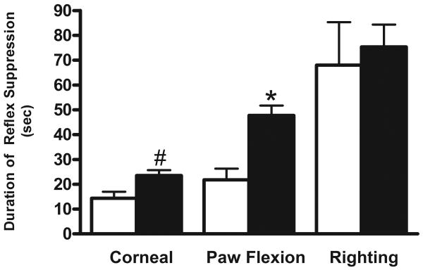
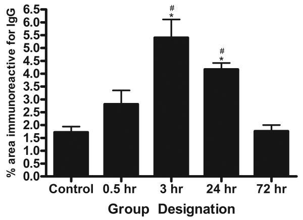
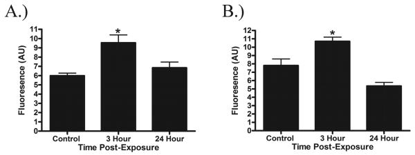
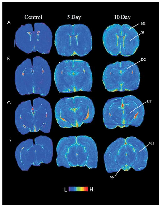
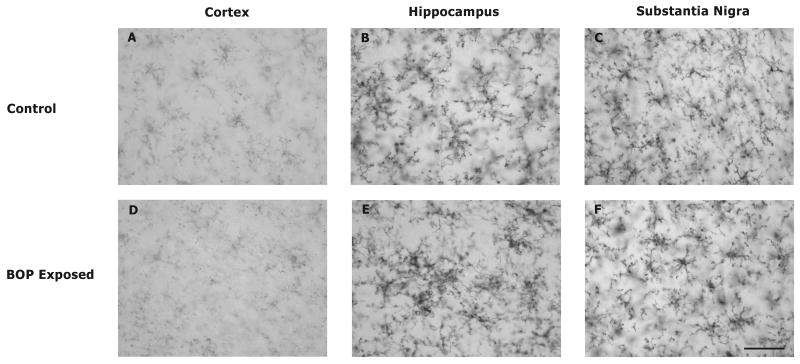
Traumatic brain injury (TBI) as a consequence of exposure to blast is increasingly prevalent in military populations, with the underlying pathophysiological mechanisms mostly unknown. In the present study, we utilized an air-driven shock tube to investigate the effects of blast exposure (120 kPa) on rat brains. Immediately following exposure to blast, neurological function was reduced. BBB permeability was measured using IgG antibody and evaluating its immunoreactivity in the brain. At 3 and 24 hr postexposure, there was a transient significant increase in IgG staining in the cortex. At 3 days postexposure, IgG immunoreactivity returned to control levels. Quantitative immunostaining was employed to determine the temporal course of brain oxidative stress following exposure to blast. Levels of 4-hydroxynonenal (4-HNE) and 3-nitrotyrosine (3-NT) were significantly increased at 3 hr postexposure and returned to control levels at 24 hr postexposure. The response of microglia to blast exposure was determined by autoradiographic localization of (3) H-PK11195 binding. At 5 days postexposure, increased binding was observed in the contralateral and ipsilateral dentate gyrus. These regions also displayed increased binding at 10 days postexposure; in addition to these regions there was increased binding in the contralateral ventral hippocampus and substantia nigra at this time point. By using antibodies against CD11b/c, microglia morphology characteristic of activated microglia was observed in the hippocampus and substantia nigra of animals exposed to blast. These results indicate that BBB breakdown, oxidative stress, and microglia activation likely play a role in the neuropathology associated with TBI as a result of blast exposure.
Copyright © 2010 Wiley-Liss, Inc.
Figures
Similar articles
-
Structural and biochemical abnormalities in the absence of acute deficits in mild primary blast-induced head trauma.J Neurosurg. 2016 Mar;124(3):675-86. doi: 10.3171/2015.1.JNS141571. Epub 2015 Aug 21. J Neurosurg. 2016. PMID: 26295915 Free PMC article.
-
Effects of Exposure to Blast Overpressure on Intracranial Pressure and Blood-Brain Barrier Permeability in a Rat Model.PLoS One. 2016 Dec 1;11(12):e0167510. doi: 10.1371/journal.pone.0167510. eCollection 2016. PLoS One. 2016. PMID: 27907158 Free PMC article.
-
Blast-Associated Shock Waves Result in Increased Brain Vascular Leakage and Elevated ROS Levels in a Rat Model of Traumatic Brain Injury.PLoS One. 2015 May 29;10(5):e0127971. doi: 10.1371/journal.pone.0127971. eCollection 2015. PLoS One. 2015. PMID: 26024446 Free PMC article.
-
Distribution of blood-brain barrier disruption in primary blast injury.Ann Biomed Eng. 2013 Oct;41(10):2206-14. doi: 10.1007/s10439-013-0805-7. Epub 2013 Apr 9. Ann Biomed Eng. 2013. PMID: 23568152
-
Blast-induced temporal alterations in blood-brain barrier properties in a rodent model.Sci Rep. 2021 Mar 15;11(1):5906. doi: 10.1038/s41598-021-84730-8. Sci Rep. 2021. PMID: 33723300 Free PMC article.
Cited by
-
What's New in Traumatic Brain Injury: Update on Tracking, Monitoring and Treatment.Int J Mol Sci. 2015 May 26;16(6):11903-65. doi: 10.3390/ijms160611903. Int J Mol Sci. 2015. PMID: 26016501 Free PMC article. Review.
-
Animal models of traumatic brain injury.Handb Clin Neurol. 2015;127:115-28. doi: 10.1016/B978-0-444-52892-6.00008-8. Handb Clin Neurol. 2015. PMID: 25702213 Free PMC article. Review.
-
A Distinct Metabolite Signature in Military Personnel Exposed to Repetitive Low-Level Blasts.Front Neurol. 2022 Apr 7;13:831792. doi: 10.3389/fneur.2022.831792. eCollection 2022. Front Neurol. 2022. PMID: 35463119 Free PMC article.
-
Novel, thalidomide-like, non-cereblon binding drug tetrafluorobornylphthalimide mitigates inflammation and brain injury.J Biomed Sci. 2023 Mar 6;30(1):16. doi: 10.1186/s12929-023-00907-5. J Biomed Sci. 2023. PMID: 36872339 Free PMC article.
-
Blast-induced axonal degeneration in the rat cerebellum in the absence of head movement.Sci Rep. 2022 Jan 7;12(1):143. doi: 10.1038/s41598-021-03744-4. Sci Rep. 2022. PMID: 34996954 Free PMC article.
References
-
- Agoston DV, Gyorgy A, Eidelman O, Pollard HB. Proteomic biomarkers for blast neurotrauma: targeting cerebral edema, inflammation, and neuronal death cascades. Journal of Neurotrauma. 2009;26:901–911. - PubMed
-
- Armonda RA, Bell RS, Vo AH, Ling G, DeGraba TJ, Crandall B, Ecklund J, Campbell WW. Wartime traumatic cerebral vasospasm: recent review of combat casualties. Neurosurgery. 2006;59:1215–1225. discussion 1225. - PubMed
-
- Banati RB, Myers R, Kreutzberg GW. PK (‘peripheral benzodiazepine’)--binding sites in the CNS indicate early and discrete brain lesions: microautoradiographic detection of [3H]PK11195 binding to activated microglia. Journal of Neurocyto. 1997;26:77–82. - PubMed
-
- Bauman RA, Elsayed N, Petras JM, Widholm J. Exposure to sublethal blast overpressure reduces the food intake and exercise performance of rats. Toxicology. 1997;121:65–79. - PubMed
-
- Beaumont A, Marmarou A, Hayasaki K, Barzo P, Fatouros P, Corwin F, Marmarou C, Dunbar J. The permissive nature of blood brain barrier (BBB) opening in edema formation following traumatic brain injury. Acta Neurochir Suppl. 2000;76:125–129. - PubMed
Publication types
MeSH terms
Grants and funding
LinkOut - more resources
Full Text Sources
Other Literature Sources
Research Materials
