

Axon growth and guidance genes identify nascent, immature, and mature olfactory sensory neurons
- PMID: 20882566
- PMCID: PMC5039016
- DOI: 10.1002/jnr.22497
Axon growth and guidance genes identify nascent, immature, and mature olfactory sensory neurons
Abstract
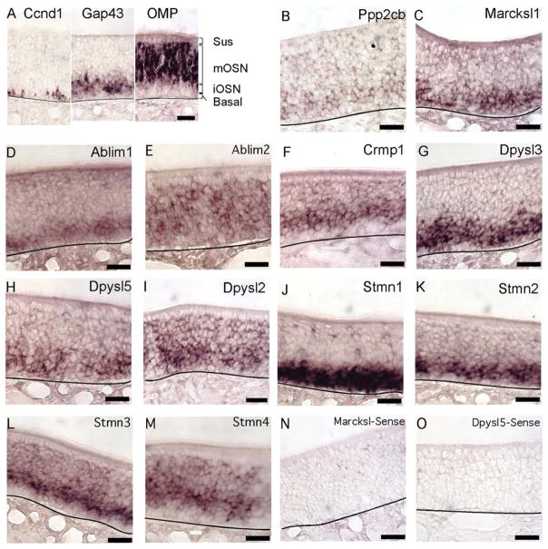
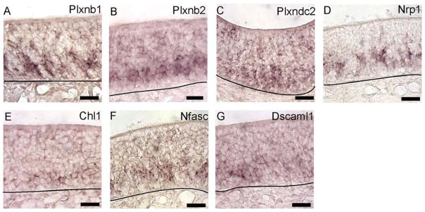
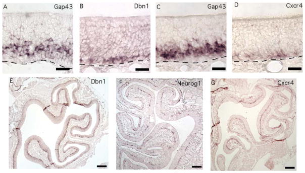
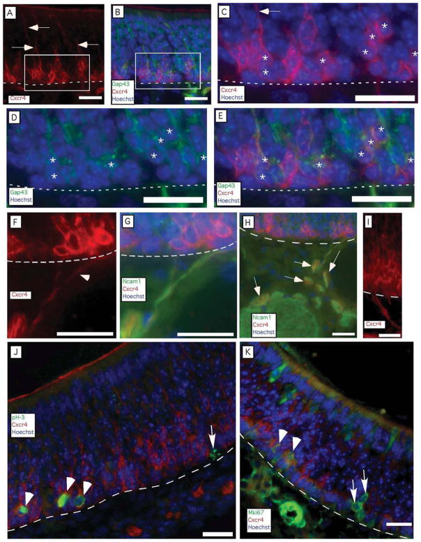
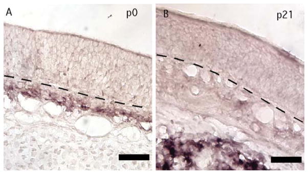
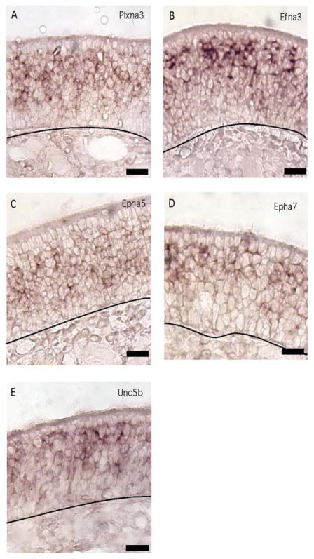
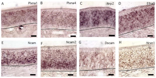
Neurogenesis of projection neurons requires that axons be initiated, extended, and connected. Differences in the expression of axon growth and guidance genes must drive these events, but comprehensively characterizing these differences in a single neuronal type has not been accomplished. Guided by a catalog of gene expression in olfactory sensory neurons (OSNs), in situ hybridization and immunohistochemistry revealed that Cxcr4 and Dbn1, two axon initiation genes, marked the developmental transition from basal progenitor cells to immature OSNs in the olfactory epithelium. The CXCR4 immunoreactivity of these nascent OSNs overlapped partially with markers of proliferation of basal progenitor cells and partially with immunoreactivity for GAP43, the canonical marker of immature OSNs. Intracellular guidance cue signaling transcripts Ablim1, Crmp1, Dypsl2, Dpysl3, Dpysl5, Gap43, Marcskl1, and Stmn1-4 were specific to, or much more abundant in, the immature OSN layer. Receptors that mediate axonal inhibition or repulsion tended to be expressed in both immature and mature OSNs (Plxna1, Plxna4, Nrp2, Efna5) or specifically in mature OSNs (Plxna3, Unc5b, Efna3, Epha5, Epha7), although some were specific to immature OSNs (Plxnb1, Plxnb2, Plxdc2, Nrp1). Cell adhesion molecules were expressed either by both immature and mature OSNs (Dscam, Ncam1, Ncam2, Nrxn1) or solely by immature OSNs (Chl1, Nfasc1, Dscaml1). Given the loss of intracellular signaling protein expression, the continued expression of guidance cue receptors in mature OSNs is consistent with a change in the role of these receptors, perhaps to sending signals back to the cell body and nucleus.
Figures
Similar articles
-
Molecular events in the cell types of the olfactory epithelium during adult neurogenesis.Mol Brain. 2013 Nov 22;6:49. doi: 10.1186/1756-6606-6-49. Mol Brain. 2013. PMID: 24267470 Free PMC article.
-
Coordination of olfactory receptor choice with guidance receptor expression and function in olfactory sensory neurons.PLoS Genet. 2018 Jan 31;14(1):e1007164. doi: 10.1371/journal.pgen.1007164. eCollection 2018 Jan. PLoS Genet. 2018. PMID: 29385124 Free PMC article.
-
Maturation of the Olfactory Sensory Neuron and Its Cilia.Chem Senses. 2020 Dec 5;45(9):805-822. doi: 10.1093/chemse/bjaa070. Chem Senses. 2020. PMID: 33075817 Free PMC article. Review.
-
Genomics of mature and immature olfactory sensory neurons.J Comp Neurol. 2012 Aug 15;520(12):2608-29. doi: 10.1002/cne.23052. J Comp Neurol. 2012. PMID: 22252456 Free PMC article.
-
Transcriptional regulation of neurogenesis in the olfactory epithelium.Cell Mol Neurobiol. 2006 Jul-Aug;26(4-6):803-21. doi: 10.1007/s10571-006-9058-4. Epub 2006 May 18. Cell Mol Neurobiol. 2006. PMID: 16708285 Free PMC article. Review.
Cited by
-
A Conserved MicroRNA Regulatory Circuit Is Differentially Controlled during Limb/Appendage Regeneration.PLoS One. 2016 Jun 29;11(6):e0157106. doi: 10.1371/journal.pone.0157106. eCollection 2016. PLoS One. 2016. PMID: 27355827 Free PMC article.
-
The cellular prion protein promotes neuronal regeneration after acute nasotoxic injury.Prion. 2020 Dec;14(1):31-41. doi: 10.1080/19336896.2020.1714373. Prion. 2020. PMID: 31950869 Free PMC article.
-
CXCL12 signaling in the development of the nervous system.J Neuroimmune Pharmacol. 2012 Dec;7(4):820-34. doi: 10.1007/s11481-011-9336-x. Epub 2012 Jan 21. J Neuroimmune Pharmacol. 2012. PMID: 22270883 Free PMC article. Review.
-
Low survival rate of young adult-born olfactory sensory neurons in the undamaged mouse olfactory epithelium.J Bioenerg Biomembr. 2019 Feb;51(1):41-51. doi: 10.1007/s10863-018-9774-8. Epub 2018 Oct 9. J Bioenerg Biomembr. 2019. PMID: 30302619 Free PMC article.
-
Molecular events in the cell types of the olfactory epithelium during adult neurogenesis.Mol Brain. 2013 Nov 22;6:49. doi: 10.1186/1756-6606-6-49. Mol Brain. 2013. PMID: 24267470 Free PMC article.
References
-
- Abercrombie M. Estimation of nuclear population from microtome sections. Anat Rec. 1946;94:239–247. - PubMed
-
- Akins MR, Greer CA. Axon behavior in the olfactory nerve reflects the involvement of catenin-cadherin mediated adhesion. J Comp Neurol. 2006;499:979–989. - PubMed
-
- Astic L, Pellier-Monnin V, Saucier D, Charrier C, Mehlen P. Expression of netrin-1 and netrin-1 receptor, DCC, in the rat olfactory nerve pathway during development and axonal regeneration. Neuroscience. 2002;109:643–656. - PubMed
-
- Au WW, Treloar HB, Greer CA. Sublaminar organization of the mouse olfactory bulb nerve layer. J Comp Neurol. 2002;446:68–80. - PubMed
Publication types
MeSH terms
Substances
Grants and funding
LinkOut - more resources
Full Text Sources
Other Literature Sources
Molecular Biology Databases
Research Materials
Miscellaneous
