. 2010 Nov;239(11):3086-97.
doi: 10.1002/dvdy.22428.

Analysis of the Hand1 cell lineage reveals novel contributions to cardiovascular, neural crest, extra-embryonic, and lateral mesoderm derivatives

Affiliations

Analysis of the Hand1 cell lineage reveals novel contributions to cardiovascular, neural crest, extra-embryonic, and lateral mesoderm derivatives

Ralston M Barnes et al. Dev Dyn. 2010 Nov.

Abstract

The basic Helix-Loop-Helix (bHLH) transcription factors Hand1 and Hand2 play critical roles in the development of multiple organ systems during embryogenesis. The dynamic expression patterns of these two factors within developing tissues obfuscate their respective unique and redundant organogenic functions. To define cell lineages potentially dependent upon Hand gene expression, we generated a mutant allele in which the coding region of Hand1 is replaced by Cre recombinase. Subsequent Cre-mediated activation of β-galactosidase or eYFP reporter alleles enabled lineage trace analyses that clearly define the fate of Hand1-expressing cells. Hand1-driven Cre marks specific lineages within the extra embryonic tissues, placenta, sympathetic nervous system, limbs, jaw, and several cell types within the cardiovascular system. Comparisons between Hand1 expression and Hand1-lineage greatly refine our understanding of its dynamic spatial-temporal expression domains and raise the possibility of novel Hand1 functions in structures not thought to be Hand1-dependent.


Figures

Figure 1
Figure 1
Generation and validation of the Hand1eGFPCre mouse line. A) The Hand1 locus is shown at the top with non-coding and coding regions indicated by the white and numbered-black boxes, respectively. The targeting vector is shown below with the dotted lines indicating regions of sequence identity between the targeting arms. The targeting strategy introduced an eGFPCre cassette into the 5’ untranslated region of Hand1, followed by an FRT-flanked PGK-neo. Homologous recombination resulted in deletion of the first exon of Hand1 and insertion of the eGFPCre gene under control of the endogenous promoter. Following germline transmission of the mutation, the PGK-neo was deleted by subsequent mating with the FLPeR mouse line. B) Southern blot of Hand1eGFPCre+Neo/+ offspring using an external probe to detect the targeted allele and C) using an internal probe to confirm the absence of transgenic incorporation of the targeting construct. D) Southern blot of Hand1eGFPCreΔNeo/+ offspring following Flp Recombinase mediated deletion of the PGK-neo. (E, F) RNA in situ hybridization on E13.5 Hand1eGFPCreΔNeo/+ embryos shows identical tissue-specific Hand1 and eGFPCre expression.
Figure 2
Figure 2
The Hand1-Lineage contributes to a subset of the first heart field and epicardium. DIG-labeled whole-mount (A, B) and transverse section (G, H, O) in situ hybridization for Hand1 on wild-type embryos at E9.5 and E10.5. LacZ staining of the Hand1LacZ (C, D, I, J, P) and Hand1eGFPCre lineage (E, F, K, L, Q). Boxed regions are shown in magnification (O,P,Q). Black Arrow denotes the left ventricle while the red arrow denotes to the epicardium. ep, epicardium; lm, lateral mesoderm; lv, left ventricle; pc, pericardium; rv, right ventricle.
Figure 3
Figure 3
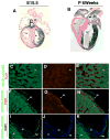
Analysis of the adult Hand1 lineage shows restriction to specific cardiovascular lineage subsets. LacZ staining of Hand1-lineage cryosections (A, B). E13.5 immunohistochemistry for Flk1 (red; D, G) and the Hand1-lineage (green; C, F). Overlay of Flk1 and the Hand1-lineage (E, H). E13.5 immunohistochemistry for the Hand1-lineage (green; I) counterstained with DAPI (blue; J). Overlay for DAPI and the Hand1-lineage (K). at, atria; da, dorsal aorta; ep, epicardium; lv, left ventricle; pt, pulmonary trunk; rv, right ventricle.
Figure 4
Figure 4
The Hand1-lineage marks discrete neural crest cell populations. Whole-mount LacZ staining of Hand1LacZ (A,J) and the Hand1-lineage (K). Transverse sections of whole mount LacZ stained Hand1LacZ (F) and Hand1-lineage (G) embryos. LacZ staining of Hand1LacZ (B, C, E, F, L, M, P, and Q), Hand1-lineage (D, G, N, and R), and Wnt1-lineage (O,S) cryosections. en, enteric; hg, hindgut; ma, mandible; mc, meckel’s cartilage; mg, midgut; mp, mandibular process; oft, outflow tract; pv, posterior vein; sg, sympathetic ganglia; t, tongue.
Figure 5
Figure 5
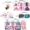
Analysis of the Hand1 limb lineage. LacZ Staining of Hand1LacZ and the Hand1 lineage at E9.5 (A–D), E10.5 (E–H), E12.5 (I–L), and E14.5 (M–P). Transverse sections of LacZ stained embryos showing fore and hind limbs (A–H). Whole-mount LacZ staining (I–P). d, dorsal; lm, lateral mesoderm; v, ventral.
Figure 6
Figure 6
The Hand1 lineage contributes to the EEM and Trophoblasts during placental development. LacZ staining of Hand1-lineage cryosections (A–H). Immunohistochemistry for the Hand1-lineage (green; I, J, M, N), Flk1 (red; K), αSM-Actin (red; O). Overlay for the Hand1 lineage, Flk1, and DAPI (blue; L). Overlay for the Hand1 lineage, and αSM-Actin, DAPI (blue; P). al, allantois; am, amnion; ch, chorion; de, decidua; ep, ectoplacental plate; gc, giant cell; lb, labyrinth; sp spongiotrophoblast; uc, umbilical chord; uv, umbilical vessels; ys, yolk sack; wj, Wharton’s jelly.
Figure 7
Figure 7
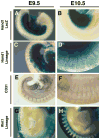
Lateral mesodermal derivatives of the Hand1 lineage gives rise to intussuceptive microvascular growth. Whole-mount LacZ staining of the Hand1LacZ (A, B), the Hand1-lineage (C, D), and the Wnt1-lineage (G, H). Whole-mount antibody staining for CD31 (E, F).
Figure 8
Figure 8
Lateral mesodermal derivatives of the Hand1 lineage give rise to endothelial progenitors of the dorsal aorta. Whole-mount LacZ staining of the Hand1-lineage (A, C). Transverse sections of whole mount LacZ stained Hand1-lineage embryos (B, D). Immunohistochemistry for the Hand1-lineage (green; F, J), Flk1 (red; G, K), and DAPI nuclear staining (blue; E, I). Overlay for the Hand1-lineage, Flk1, and DAPI (H, L). cv, cardinal vein; da, dorsal aorta; eem, extraembryonic mesoderm; lm, lateral mesoderm; sg, sympathetic ganglia.

References

    1. Abe M, Tamamura Y, Yamagishi H, Maeda T, Kato J, Tabata MJ, Srivastava D, Wakisaka S, Kurisu K. Tooth-type specific expression of dhand/HAND2: possible involvement in murine lower incisor morphogenesis. Cell Tissue Res. 2002;310:201–212. - PubMed
    1. Ahn K, Mishina Y, Hanks MC, Behringer RR, Crenshaw EB., 3rd BMPR-IA signaling is required for the formation of the apical ectodermal ridge and dorsal-ventral patterning of the limb. Development. 2001;128:4449–4461. - PubMed
    1. Barbosa AC, Funato N, Chapman S, McKee MD, Richardson JA, Olson EN, Yanagisawa H. Hand transcription factors cooperatively regulate development of the distal midline mesenchyme. Developmental Biology. 2007;310:154–168. - PMC - PubMed
    1. Barnes RM, Firulli AB. A Twist of insight, the role of Twist-Family bHLH factors in development. Int J Dev Biol. 2009;53:909–924. - PMC - PubMed
    1. Chen H, Johnson RL. Interactions between dorsal-ventral patterning genes lmx1b, engrailed-1 and wnt-7a in the vertebrate limb. Int J Dev Biol. 2002;46:937–941. - PubMed

Publication types

Substances

LinkOut - more resources