Comparative Study
. 2010:10:132.
doi: 10.1673/031.010.13201.

Study of acrosome formation, interspecific and intraspecific, in the testicular lobes of some pentatomid species

Affiliations
Comparative Study

Study of acrosome formation, interspecific and intraspecific, in the testicular lobes of some pentatomid species

Hederson V Souza et al. J Insect Sci. 2010.

Abstract

The objective was to compare the formation of the acrosome in the testicular lobes of the species Antiteuchus tripterus F. and Platycarenus umbractulatus F., belonging to the subfamily Discocephalinae, and Euschistus heros F., Mormidea quinqueluteum L., Oebalus sp. and Thyanta perditor F., belonging to the Pentatominae (Heteroptera: Pentatomidae). It was found that, in general, the behavior of periodic acid Schiff-positive granules for all of the species analyzed is similar for these species. In the beginning of spermiogenesis, there is a central granule that migrates to one of the extremities of the spermatid, and later, it becomes elongated and cannot be distinguished in the spermatozoa. Some species such as A. tripterus, E. heros and P. umbractulatus showed significant differences in the behavior of the PAS-positive granule in certain lobes, suggesting the formation of spermatozoa with non-fertile functions.


Figures

Figure 1.
Figure 1.
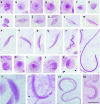
Cells of the testicular lobes of Antiteuchus tripterus stained with PAS. a–n - Cells of lobes 1–3; a, b - round spermatid evidencing PAS-positive granules dispersed throughout the cell; (c–h) spermatid in elongation with PAS-positive reaction located in the periphery of the cell (c–g) or in the center (h); i–n - spermatids in the final stage of development without PAS-positive reaction; o, p - round spermatids of lobe 4 with intense PAS-positive reaction; q, r round spermatid of lobe 6 with PAS-positive reaction in the center; s–z - cells of lobe 5; s, t - round spermatid: note the different sizes of the cells and PAS-positive reaction; u–x - large spermatids (u), medium (v) and small (x); z- spermatid in the final stage of development. Bar= 10 µm. High quality figures are available online.
Figure 2.
Figure 2.
Cells of the testicular lobes of Euschistus heros stained with PAS. a–s - Cells of lobes 1–3; a–e - round spermatid evidencing in (a) several dispersed PAS-positive granules in the cell or with the PAS-positive reaction in the center of the cell (c, e); h–q - medium spermatids with PAS-positive granules moved to one of the extremities of the cell (h–k) and later they are elongated towards the center (l–q); r, s - spermatids in the final stage of development evidencing PAS-positive reaction in whole cell; t, u - round spermatids of lobe 5 evidencing strong PAS-positive reaction; v–y - cells of lobes 4 and 6 showing in (v–z) round spermatids with intense PAS-positive reaction moved to periphery; w, y - spermatids in the final stage of development with PAS- positive reaction in the inferior head region; a1, b1 - spermatid in the final stage of development of lobe 5 with a wide PAS-positive reaction throughout the cell. Bar= 10 µm. High quality figures are available online.
Figure 3.
Figure 3.
Cells of the testicular lobes of Mormidea quinqueluteum stained with PAS. a–i - Round spermatids evidencing in (a, b) several dispersed PAS-positive granules in the cell, in (c, d) the PAS-positive reaction is located in the center of the cell and moved to the periphery (e, f), acquiring the C shape (g–i); j–p - medium spermatids with PAS- positive granule at one of the extremities (j, k), which is elongated toward the cell center (m) or is at the periphery of the cell (n–p); q, r spermatids at the end of elongation evidencing a continuous PAS-positive reaction in whole head. Bar= 10 µ/m. High quality figures are available online.
Figure 4.
Figure 4.
Cells of the testicular lobes of Oebalus sp. stained with PAS. a–f - Round spermatids with several dispersed PAS-positive granules in the cell (a, c), moved to the periphery (d, f); g–k - spermatids in the beginning of development with PAS-positive reaction at one of the extremities of the cell; l–n - medium spermatids with PAS-positive granule being elongated towards other extremity of the cell; o–r - spermatids in the final stage of development acquiring the C shape (g–i); j–p - medium spermatids evidencing a continuous PAS-positive reaction in whole head. Bar= 10 µm. High quality figures are available online.
Figure 5.
Figure 5.
Cells of the testicular lobes of Platycarenus umbractulatus stained with PAS. a– m - Cells of lobes 1–3 and 7; a–g round spermatids evidencing in (a, b) several dispersed PAS-positive granules in the cell, in (c, d) the PAS-positive reaction is located in the center of the cell, moved to the periphery forming a C shape (e–g); h–k - medium spermatids with PASpositive reaction on one on the sides; l–m - spermatids in the final stage of development with PAS-positive reaction in whole head, n–r - Cells of lobes 4 and 6; n, o - spermatids in elongation with PAS-positive reaction forming a C shape, p, q medium spermatids with PAS-positive granules throughout the cell, more evident at one of the extremities, r spermatid in the final stage of development with a PAS- positive reaction at one of the extremities, s–e1 - Cells of testicular lobe 5; s–x round spermatids with different PAS-positive reaction, z–el - spermatids in development with different PAS-positive reaction. Bar= 10 µm. High quality figures are available online.
Figure 6.
Figure 6.
Cells of the testicular lobes of Thyanta perditor stained with PAS. a–f - Round spermatids evidencing PAS-positive reaction located in the center of the cell; g–i - the PAS-positive reaction remains in the center of the cell until the final stage of elongation of the spermatid; j - elongated spermatid demonstrating a notable elongation of the central PAS-positive reaction; k, l - spermatids in the final stage of development with continuous PAS-positive reaction in the whole head. Bar= 10 µm. High quality figures are available online.

Similar articles

Cited by

References

    1. Anderson WA, Personne P. The form and function of spermatozoa: a comparative view. In: Afzelius BA, editor. The Functional Anatomy of the Spermatozoon. Pergamon Press; 1975. pp. 3–14.
    1. Baccetti B. Insect sperm cells. In: Treherne JE, Berridge MJ, Wigglesworth VB, editors. Advances in Insect Physiology 9. Academic Press; 1972. pp. 315–397.
    1. Bowen RH. Notes on the occurrence of abnormal mitosis in spermatogenesis. Biological Bulletin. 1922;43:184–203.
    1. Bowen RH. On the acrosome of the animal sperm. Anatomical Record. 1924;28:1–13.
    1. Garcia A. Manual de técnicas citogenéticas. Colégio de Pósgraduados; Chapingo, México: 1990.

Publication types