. 2011 May;44(3):219-28.
doi: 10.3109/08916934.2010.519746. Epub 2010 Oct 1.

Increased T regulatory cells lead to development of Th2 immune response in male SJL mice

Affiliations

Increased T regulatory cells lead to development of Th2 immune response in male SJL mice

Shabbir Hussain et al. Autoimmunity. 2011 May.

Abstract

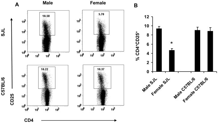
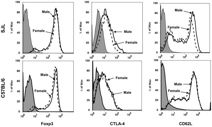
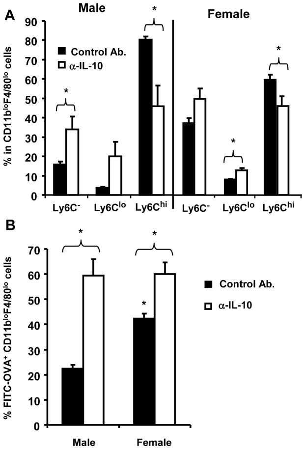
SJL mice represent a mouse model in which young adult females are susceptible to autoimmune disease, while age-matched males are relatively resistant. T cells primed in female SJL mice secrete cytokines associated with a Th1 phenotype. By contrast, T cells primed in males secrete cytokines associated with a Th2 phenotype. Activation of Th2-type T cells in males vs. Th1 cells in females correlates with increased CD4(+)CD25(+) T regulatory cells (Treg) in males. T cells primed in males depleted of CD4(+)CD25(+) T cells preferentially secrete IFN-γ and decreased IL-4 and IL-10 compared to CD4(+)CD25(+) T-cell-sufficient males, suggesting that Treg influence subsequent antigen-specific cytokine secretion. Treg from males and females exhibit equivalent in vitro T-cell suppression. Treg from males express increased CTLA-4 and CD62L and preferentially secrete IL-10. These data suggest that an increased frequency of IL-10 secreting Treg in male SJL mice may contribute resistance to autoimmune disease by favoring the development of Th2 immune responses.


Figures

Figure 1
Figure 1. Female SJL mice are deficient in CD4+CD25+ T cells
Spleen cells from age-matched male and female SJL and C57BL/6 mice were analyzed by flow cytometry as described in Materials and Methods. Male and female SJL and C57BL/6 mice were compared for CD25 expression on CD4+ T cells. Data are representative of 4–8 independent experiments with similar results (A) and mean ± SEM (B). *p < 0.001 compared to SJL males and C57BL/6 males and females.
Figure 2
Figure 2. Treg in female SJL mice express reduced CTLA-4 and CD62L
CD4+CD25+ T cells from age-matched male and female SJL and C57BL/6 mice were compared for the expression of Foxp3, CTLA-4 and CD62L by flow cytometry. Filled histograms represent isotype control, dashed lines represent data from females and continuous lines represent data from males. Data are representative of 4-8 independent experiments with similar results.
Figure 3
Figure 3. Reduced thymic maturation of CD4+Foxp3+ cells in female SJL mice
Male and female SJL and C57BL/6 mice were compared for the expression of thymic Foxp3 within single positive CD4 T cells. Numbers represent percentages of Foxp3+ cells. Data presented are representative of 3 independent experiments with similar results (A) and mean ± SEM (B). *p < 0.05 compared to SJL males and C57BL/6 males and females.
Figure 4
Figure 4. Phenotypic characterization of Foxp3+ T cells in spleens of male and female SJL mice
Spleen cells from age-matched male and female SJL mice were analyzed by flow cytometry as described in Materials and Methods. (A) Foxp3 expression was compared between male and female SJL CD4+ T cells. (B) Expression of CD25, CTLA-4, CD62L, GITR, CD103 and TNFR2 was compared on Foxp3+CD4+ T cells from male and female SJL spleens. Filled histograms represent isotype control, dotted lines represent data from females and continuous lines represent data from males. Data are representative of spleens from 4 mice per group processed individually.
Figure 5
Figure 5. Equivalent suppression by Treg from male and female SJL mice
CD4+CD25+ Treg and CD4+CD25 T cells were purified from spleens and lymph nodes of naïve male and female SJL mice. CD4+CD25 T cells (T effector) were labeled with CFSE and activated with anti-CD3 in presence of irradiated spleen cells. Treg were added at the indicated ratios and proliferation analyzed by flow cytometry at 72 h. Data are representative of 3 experiments.
Figure 6
Figure 6. Treg from male SJL mice secrete increased IL-10
CD4+CD25+ Treg from spleens of naïve 6 wk-old males and females were stimulated with anti-CD3 for 48 h. IL-10 concentrations determined by ELISA. Data are representative of 3 experiments performed in triplicate. Error bars = SEM. *p < 0.05 compared to females.
Figure 7
Figure 7. Treg regulate T cell priming in male SJL mice
Male SLJ mice were depleted of CD4+CD25+ T cells prior to immunization and challenged with antigen 5 days post-immunization. Draining lymph nodes removed 24 h later. Cytokines secretion at 48 h after antigen stimulation were determined by ELISA. Results are the average of at least 3 experiments with 2 mice per group. Error bars = SEM. *p ≤ 0.01 compared to controls.

Similar articles

Cited by

References

    1. Tang Q, Bluestone JA. The Foxp3+ regulatory T cell: a jack of all trades, master of regulation. Nat Immunol. 2008;9:239–44. - PMC - PubMed
    1. Brusko TM, Putnam AL, Bluestone JA. Human regulatory T cells: role in autoimmune disease and therapeutic opportunities. Immunol Rev. 2008;223:371–90. - PubMed
    1. Belkaid Y. Role of Foxp3-positive regulatory T cells during infection. Eur J Immunol. 2008;38:918–21. - PMC - PubMed
    1. Van Wijk F, Roord ST, Vastert B, De Kleer I, Wulffraat N, Prakken BJ. Regulatory T cells in autologous stem cell transplantation for autoimmune disease. Autoimmunity. 2008;41:585–91. - PubMed
    1. Houot R, Perrot I, Garcia E, Durand I, Lebecque S. Human CD4+CD25high regulatory T cells modulate myeloid but not plasmacytoid dendritic cells activation. J Immunol. 2006;176:5293–8. - PubMed

Publication types