. 2010 Oct;23(5):497-508.
doi: 10.1089/vim.2010.0043.

Synthetic antigens representing the antigenic variation of human hepatitis C virus

Affiliations

Synthetic antigens representing the antigenic variation of human hepatitis C virus

Kyung Hee Kang et al. Viral Immunol. 2010 Oct.

Abstract

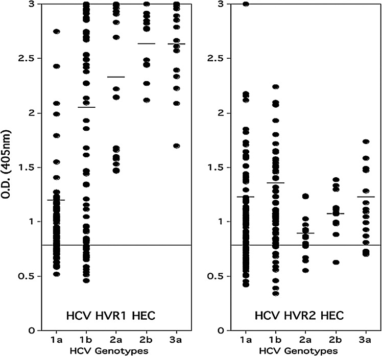
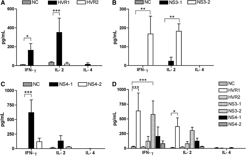
Immune responses against hepatitis C virus (HCV) have been studied by numerous groups. However, details concerning the production of antibodies to antigenically variable epitopes remain to be elucidated. Since the sequences of the variable regions of several HCV proteins are different among the virus strains infecting patients, we decided to design peptide combinations that represent the theoretical maximum antigenic variation of each epitope to be used as capture antigens. We prepared six peptide mixtures (hypervariable epitope constructs; HECs) representing six different epitopes from structural and non-structural proteins of HCV from genotypes 1-6. Plasma from 300 HCV patients was tested to determine if their antibodies recognize the synthetic constructs. All the patients were chronically infected with diverse HCV genotypes and did not receive antiviral treatment. Antibodies to one or more of the HECs were detected in all of the HCV-infected individuals. Immunogenicity of the HCV HECs was also evaluated in outbred and inbred mice. Strong HEC-specific antibodies were produced, and cellular responses were also induced that were Th-1 rather than Th-2. Our results show that HCV HECs are both antigens that can be used to detect the broad cross-reactivity of antibodies from HCV-infected patients, and strong immunogens that can induce antigen-specific humoral and cellular immune responses in mice.


Figures

FIG. 1.
FIG. 1.
Relative antibody titers to the HVR1 and HVR2 HECs in plasma of HCV-positive patients (group 1). Dot plot showing relative OD values obtained when individual plasma samples from HCV patients were tested for antibodies to HVR1 and HVR2 HECs. All sera were diluted 1:100. The samples were considered positive if their optical density at 405 nm was more than twice that of the negative sera. The horizontal line indicates the OD value that is twice the average of four HCV-negative serum samples. The sera did not react with a negative control HEC based on HIV-1 (data not shown).
FIG. 2.
FIG. 2.
Relative antibody titers to six HCV HECs in 50 plasma samples from HCV-positive patients (group 2). Recognition of HVR1, HVR2, NS3-1, NS3-2, NS4-1 and NS4-2 HCV HECs by patients infected with different genotypes of HCV (1a, 1b, 1a/b, 2b, 2a/c, 3a, and 4c/d). Each dot represents an individual plasma sample from an HCV patient. Antibody end-point titers for binding to HCV HEC were determined based on the OD value of normal human sera used as negative control.
FIG. 3.
FIG. 3.
Immunogenicity of HCV HECs in BALB/c mice. (A) ELISA of sera from mice immunized with HVR1, HVR2, NS3-1, NS3-2, NS4-1, and NS4-2 HECs. The HCV HEC-specific antibody responses were determined by ELISA. BALB/c mice immunized with a mixture of HCV HECs produced antibodies that recognized each individual HEC. Sera were obtained 7 d after the final immunization. Serum samples obtained before immunization were used as negative controls. (B) Recognition of HCV particles (purified from plasma of patients infected by different HCV genotypes) by antibodies from mice immunized with a mixture of 6 HCV HECs. Pre-bleed sera obtained before immunization were used as negative controls. (C and D) Cross-reactivity of sera from mice immunized with HVR1, HVR2, NS3-1, NS3-2, NS4-1, and NS4-2 HECs. HVR1 (C) and HVR2 (D) analogs representing genotypes from 1a to 6a were used as capturing antigen in triplicate. Sera from three mice were evaluated with antibody dilution rates of 1:100, 1:500, 1:1000, 1:2000, 1:4000, 1:8000, 1:16,000, and 1:20,000. Data shown are the mean ± SD of the antibody titer (in 1 per dilution rate) as assessed by ELISA with serum from four individual mice and analyzed in triplicate. The data in Fig. 3A were regarded as statistically highly significant (***p < 0.001).
FIG. 4.
FIG. 4.
HCV HEC-specific lymphocyte proliferation as measured by thymidine incorporation. Six outbred mice (CFW) per group were immunized with a combination of two HECs, or with all six HECs together. The two-HEC combinations were (1) HVR1 and HVR2, (2) NS3-1 and NS3-2, and (3) NS4-1 and NS4-2. An unrelated peptide derived from SIV was used as a negative control (NC). Lymphocytes from lymph nodes were stimulated in vitro with each HCV HEC at 5 μg/mL for 3 d. Following 14 h of incubation with 1 μCi of tritiated thymidine per well, the incorporated thymidine was determined and is presented as counts per minute (cpm). (A) Mice immunized with the HVR1 and HVR2 HECs. (B) Mice immunized with the NS3-1 and NS3-2 HECs. (C) Mice immunized with the NS4-1 and NS4-2 HECs. (D) Mice immunized with all six HECs combined. Data shown are the mean ± SD of the value of incorporated [3H]thymidine from six individual mice and analyzed in triplicate. Statistical analysis was conducted using one-way ANOVA (Dunnett's multiple comparison test: *p < 0.05; **p < 0.01).
FIG. 5.
FIG. 5.
Detection of cytokines secreted from HCV HEC-specific lymphocytes. IFN-γ, IL-2, and IL-4 cytokines secreted from HEC-specific T lymphocytes were quantitated by cytokine ELISA. In each group, 5 × 105 lymphocytes from the spleens were stimulated in vitro with each HCV HEC at 5 μg/mL for 2 d, and the supernatants were collected to measure cytokine levels. An HCV-unrelated peptide (SIV) was used as a negative control (NC). (A) Mice immunized with HVR1 and HVR2 HECs. (B) Mice immunized with NS3-1 and NS3-2 HECs. (C) Mice immunized with NS4-1 and NS4-2 HECs. (D) Mice immunized with all six HECs combined. Data shown are the mean ± SD of cytokine concentration values in picograms per milliliter from three to four individual mice and analyzed in triplicate. Statistical analysis was conducted using two-way ANOVA (Bonferroni's post-test: *p < 0.05; **p < 0.01; ***p < 0.001).

References

    1. Aberle JH. Formann E. Steindl-Munda P, et al. Prospective study of viral clearance and CD4(+) T-cell response in acute hepatitis C primary infection and reinfection. J Clin Virol. 2006;36:24–31. - PubMed
    1. Anderson DE. Malley A. Benjamini E. Gardner MB. Torres JV. Hypervariable epitope constructs as a means of accounting for epitope variability. Vaccine. 1994;12:736–740. - PubMed
    1. Anderson DE. Singapuri A. Kang KH. Montefiori DC. Torres JV. Timing of retroviral infection influences anamnestic immune response in vaccinated primates. Viral Immunol. 2005;18:689–694. - PubMed
    1. Blum HE. Variants of hepatitis B, C and D viruses: molecular biology and clinical significance. Digestion. 1995;56:85–95. - PubMed
    1. Bowen DG. Walker CM. Adaptive immune responses in acute and chronic hepatitis C virus infection. Nature. 2005;436:946–952. - PubMed

LinkOut - more resources