. 2010 Dec 15;432(3):549-56.
doi: 10.1042/BJ20100936.

PP2B/calcineurin-mediated desensitization of TRPV1 does not require AKAP150

Affiliations

PP2B/calcineurin-mediated desensitization of TRPV1 does not require AKAP150

Elaine D Por et al. Biochem J. .

Abstract

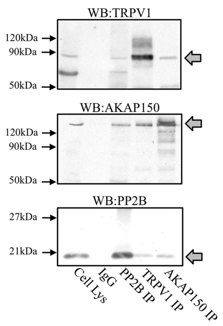
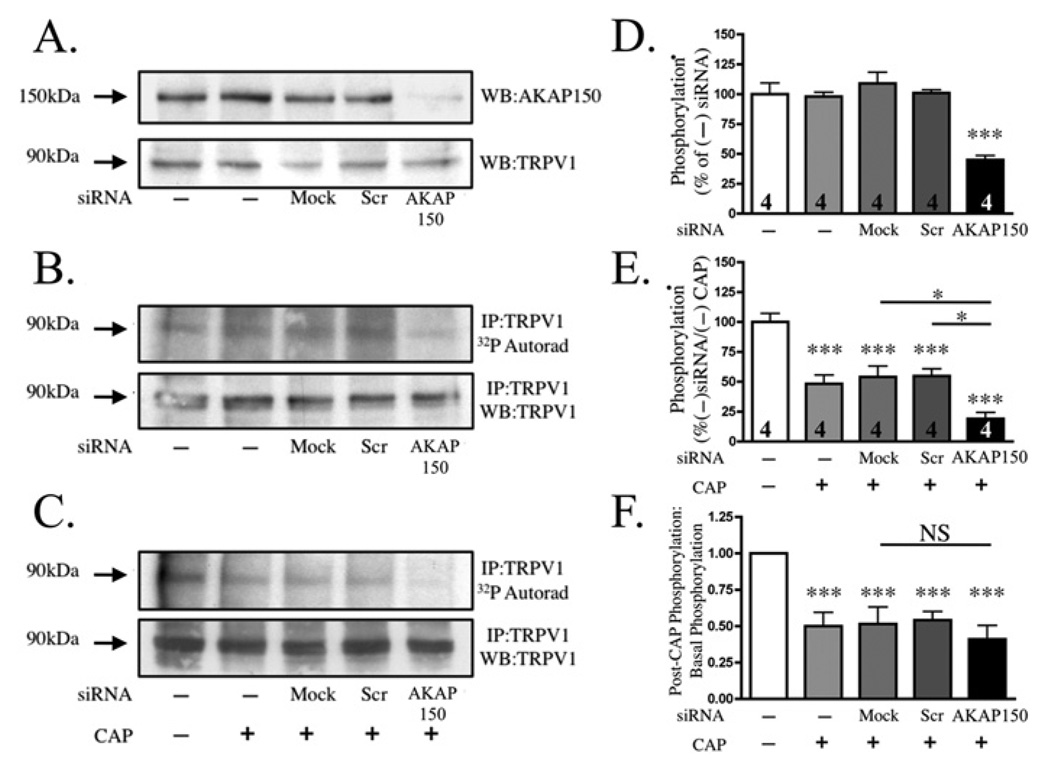
Activation of protein kinases and phosphatases at the plasma membrane often initiates agonist-dependent signalling events. In sensory neurons, AKAP150 (A-kinase-anchoring protein 150) orientates PKA (protein kinase A), PKC (protein kinase C) and the Ca2+/calmodulin-dependent PP2B (protein phosphatase 2B, also known as calcineurin) towards membrane-associated substrates. Recent evidence indicates that AKAP150-anchored PKA and PKC phosphorylate and sensitize the TRPV1 (transient receptor potential subfamily V type 1 channel, also known as the capsaicin receptor). In the present study, we explore the hypothesis that an AKAP150-associated pool of PP2B catalyses the dephosphorylation and desensitization of TRPV1. Biochemical, electrophysiological and cell-based experiments indicate that PP2B associates with AKAP150 and TRPV1 in cultured TG (trigeminal ganglia) neurons. Gene silencing of AKAP150 reduces basal phosphorylation of TRPV1. However, functional studies in neurons isolated from AKAP150-/- mice indicate that the anchoring protein is not required for pharmacological desensitization of TRPV1. Behavioural analysis of AKAP150-/- mice further support this notion, demonstrating that agonist-stimulated desensitization of TRPV1 is sensitive to PP2B inhibition and does not rely on AKAP150. These findings allow us to conclude that pharmacological desensitization of TRPV1 by PP2B may involve additional regulatory components.


Figures

Figure 1
Figure 1. AKAP150 and TRPV1 associate with the PP2B-B subunit in rat primary sensory neurons
TG neurons were lysed and incubated with antibodies specific against TRPV1, AKAP150 and PP2B-B. Western blotting (WB) was performed to detect co-immunoprecipitates (IP) and expression levels in plasma membrane fractions. IgG and cell lysates (Cell Lys) were assessed for specificity of the TRPV1 antibody and expression profiles of all proteins. Molecular mass markers (in kDa) are shown on the left-hand side. Arrows on the right-hand side denote immunoreactive bands of interest. Results are representative of four independent experiments.
Figure 2
Figure 2. AKAP150 does not mediate CAP-induced dephosphorylation of TRPV1 in primary sensory neurons
TG neurons were transfected with siRNA directed against AKAP150, silencer negative siRNA (Scr) or no siRNA (mock) and assessed for [32P]Pi incorporation (TRPV1 phosphorylation) by immunoprecipitation (IP) and Western blotting (WB). (A) AKAP150 expression levels in untransfected (−), mock, scrambled- and AKAP150-siRNA transfected cells. (B) Basal phosphorylation levels of TRPV1 following siRNA transfection. (C) Phosphorylation of TRPV1 following application of CAP (100 nM; 30 s). For (A–C) the molecular mass in kDa is indicated on the left-hand side. (D) Quantification of TRPV1 basal phosphorylation levels following untransfected (−), mock, scrambled- and AKAP150-siRNA transfection (significant compared with mock-transfected cells). Phosphorylation levels of TG-transfected neurons following activation (E) were normalized with basal levels of TRPV1 phosphorylation (F). *P < 0.05, **P < 0.01, and ***P < 0.005, as determined using two-way ANOVA with Bonferroni correction. Results are representative of four independent experiments.
Figure 3
Figure 3. PP2B associates with TRPV1 in the absence of AKAP150 in primary sensory neurons
TG neurons were either mock-transfected or transfected with siRNA directed against AKAP150, and treated with vehicle or CAP (100 nM, 30 s). Following treatment, crude plasma membrane homogenates were collected for each treatment condition and immunoprecipitated (IP) with antibodies specific for TRPV1. Arrows on the left-hand side denote the predicted molecular mass of the protein of interest. Results are representative of three independent experiments. WB, Western blot.
Figure 4
Figure 4. AKAP150 association with PP2B does not mediate CAP-induced desensitization of TRPV1
CHO cells transiently transfected with TRPV1, TRPV1 + AKAP150 or TRPV1 + AKAPΔPP2B were assessed for CAP-induced Ca2+ accumulation. (A) Representative traces. Following acquisition of baseline, CAP (100 nM) was administered for 30 s (shaded bar), and the cells were rinsed with SES buffer for 3 min. To assess desensitization, a second application of CAP was administered for 30 s following the 3 min rinse. Accumulation of Ca2+ was calculated from the 340/380 ratio. (B) The net change in Ca2+ (ΔF340/380) was calculated by subtracting the basal F340/380 Ca2+ level from the peak F340/380 Ca2+ level achieved, n = 74–90 neurons per transfection group. (C) Ca2+ accumulation from CAP/CAP treatments were normalized to those for vehicle/CAP treatments, and are displayed as normalized desensitization. (D) Ca2+ accumulation from CAP/CAP treatments were normalized to those for vehicle/CAP treatments following pre-treatment with vehicle (H20) or cell-permeant CAIP (100 mM, 30 min), and are displayed as normalized desensitization. ***P < 0.005 and NS = no significance, as determined by one-way ANOVA, with Bonferroni post-hoc analysis. n values are indicated in the bars of each histogram.
Figure 5
Figure 5. AKAP150 knockdown or knockout does not effect TRPV1 localization to the plasma membrane
(A) TG neurons isolated from wild-type (WT) and AKAP150−/− (KO) mice were cultured and surface biotinylated to identify plasma membrane proteins exposed to the extracellular space. (B) PKA RII subunit-binding proteins were detected in lysates generated from homogenized TGs dissected and frozen from wild-type (WT, AKAP150+/+) and AKAP150−/− (KO) mice, using 32P-radiolabelled RIIα as a probe. Control blots indicate correct expression profiles for AKAP150 and β-actin. (C) TG neurons from rats were transfected in a mock fashion or with AKAP150-specific siRNA, and homogenates were differentially centrifuged to isolate the crude plasma membrane fraction. Equal aliquots (30 µg) of plasma membrane (PM) lysates were resolved and analysed for AKAP150, TRPV1 and β1-integrin expression. The molecular mass in kDa is indicated on the left-hand side. Arrows on the right-hand side denote immunoreactive bands of interest. Results are representative of four independent experiments.
Figure 6
Figure 6. AKAP150 is not required for behavioural TRPV1 desensitization
Wild-type (AKAP150+/+) and AKAP150−/− mice were assessed for nocifensive behaviour as determined by time spent licking and flinching the injected hind-paw. For control experiments, both groups were injected with vehicle (20% NMP) followed 15 min later with CAP (0.5 µg) in a 10 µl injection volume. For desensitization experiments, both groups were injected with CAP (0.5 µg/10 µl) followed by a second injection of CAP (0.5 µg/10 µl) 15 min later. FK-506 (20 mg/10 ml) was administered prior to the first CAP injection, at the same injection site. Nocifensive behaviour was recorded over a 5 min period for all experiments. **P < 0.01 and ***P < 0.005, indicates the significance from the respective genotype vehicle/CAP treatment as measured by one-way ANOVA. n = 6–10 animals/group.
Figure 7
Figure 7. Genetic ablation of AKAP150 does not affect CAP-induced desensitization of TRPV1 activity
Sample CAP (300 nM, 30 s)-current traces from TG neurons isolated from wild-type (AKAP150+/+) (A–C) or AKAP150−/− (D–F) animals following vehicle (Veh), W-7 (500 mM) or FK-506 (10 µM) co-treatments, as indicated. The time between concurrent CAP applications is 2 min. (G) Normalized CAP-desensitization from the total neurons. Normalization of the second CAP applications were carried out against the first CAP application. *P < 0.05 and **P < 0.01 indicate significance from vehicle-treated genotype, as determined by one-way ANOVA. n values are indicated in the bars of each graph.

References

    1. Scott JD, Pawson T. Cell signaling in space and time: where proteins come together and when they’re apart. Science. 2009;326:1220–1224. - PMC - PubMed
    1. Wong W, Scott JD. AKAP signalling complexes: focal points in space and time. Nat. Rev. Mol. Cell Biol. 2004;5:959–970. - PubMed
    1. Klauck TM, Faux MC, Labudda K, Langeberg LK, Jaken S, Scott JD. Coordination of three signaling enzymes by AKAP79, a mammalian scaffold protein. Science. 1996;271:1589–1592. - PubMed
    1. Jeske NA, Diogenes A, Ruparel NB, Fehrenbacher JC, Henry M, Akopian AN, Hargreaves KM. A-kinase anchoring protein mediates TRPV1 thermal hyperalgesia through PKA phosphorylation of TRPV1. Pain. 2008;138:604–616. - PMC - PubMed
    1. Jeske NA, Patwardhan AM, Ruparel NB, Akopian AN, Shapiro MS, Henry MA. A-kinase anchoring protein 150 controls protein kinase C-mediated phosphorylation and sensitization of TRPV1. Pain. 2009;146:301–307. - PMC - PubMed

Publication types

MeSH terms