

Regulation of valvular interstitial cell phenotype and function by hyaluronic acid in 2-D and 3-D culture environments
- PMID: 20884350
- PMCID: PMC3021089
- DOI: 10.1016/j.matbio.2010.09.001
Regulation of valvular interstitial cell phenotype and function by hyaluronic acid in 2-D and 3-D culture environments
Abstract
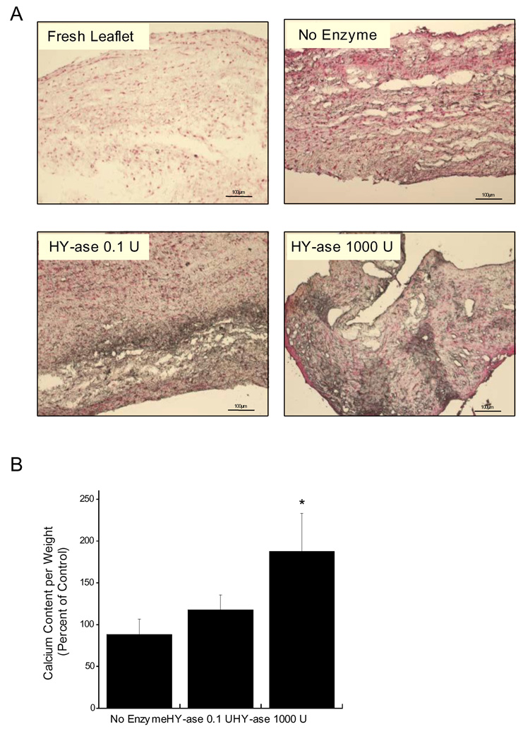
Disruption of the extracellular matrix (ECM) is frequently found in calcific aortic valve disease (CAVD), yet the role of ECM components in valvular interstitial cell (VIC) function and dysfunction remains poorly understood. This study examines the contributions of exogenous and endogenous hyaluronic acid (HA), in both two-dimensional (2-D) and 3-D environments, in regulating the phenotype and calcification of VICs. VIC calcification was first assessed in a 2-D setting in which the cells were exposed to different molecular weights of exogenous HA presented in either an immobilized or soluble form. Delivery of HA suppressed nodule formation in a molecular weight-dependent manner, while blocking VIC recognition of HA via an antibody to CD44 abolished these nodule-suppressive effects and stimulated other hallmarks of valvular dysfunction. These 2-D results were then validated in a more physiologically-relevant setting, using an approach that allowed the characterization of VIC phenotype in response to HA alterations in the native 3-D environment. In this approach, leaflet organ cultures were analyzed following treatment with anti-CD44 or with hyaluronidase to specifically remove HA. Disruption of VIC-HA interactions upregulated markers of VIC disease and induced leaflet mineralization. Similarly, HA-deficient leaflets exhibited numerous hallmarks of CAVD, including increased VIC proliferation, apoptosis, increased expression of disease-related markers, and mineralization. These findings suggest that VIC-HA interactions are crucial in maintaining a healthy VIC phenotype. Identification ECM components that can regulate VIC phenotype and function has significant implications for understanding native valve disease, investigating possible treatments, and designing new biomaterials for valve tissue engineering.
Copyright © 2010 International Society of Matrix Biology. Published by Elsevier B.V. All rights reserved.
Figures


References
-
- Aikawa E, Whittaker P, Farber M, Mendelson K, Padera RF, Aikawa M, Schoen FJ. Human semilunar cardiac valve remodeling by activated cells from fetus to adult: implications for postnatal adaptation, pathology, and tissue engineering. Circulation. 2006;113:1344–1352. - PubMed
-
- Allison DD, Braun KR, Wight TN, Grande-Allen KJ. Differential effects of exogenous and endogenous hyaluronan on contraction and strength of collagen gels. Acta. Biomater. 2009;5:1019–1026. - PubMed
-
- Barbosa I, Garcia S, Barbier-Chassefiere V, Caruelle JP, Martelly I, Papy-Garcia D. Improved and simple micro assay for sulfated glycosaminoglycans quantification in biological extracts and its use in skin and muscle tissue studies. Glycobiology. 2003;13:647–653. - PubMed
-
- Chao H, Spicer AP. Natural antisense mRNAs to hyaluronan synthase 2 inhibit hyaluronan biosynthesis and cell proliferation. J. Biol. Chem. 2005;280:27513–27522. - PubMed
Publication types
MeSH terms
Substances
Grants and funding
LinkOut - more resources
Full Text Sources
Miscellaneous
