

Mapping anterior temporal lobe language areas with fMRI: a multicenter normative study
- PMID: 20884358
- PMCID: PMC2997157
- DOI: 10.1016/j.neuroimage.2010.09.048
Mapping anterior temporal lobe language areas with fMRI: a multicenter normative study
Abstract
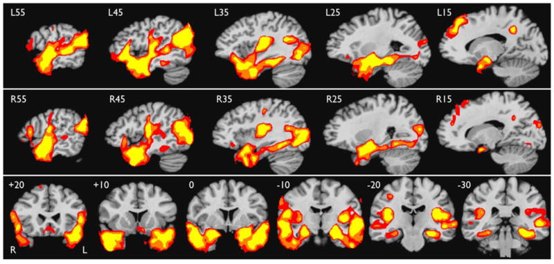
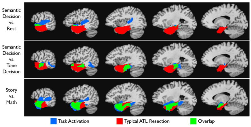
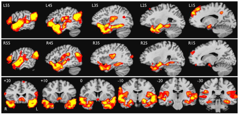
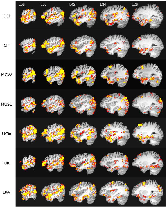
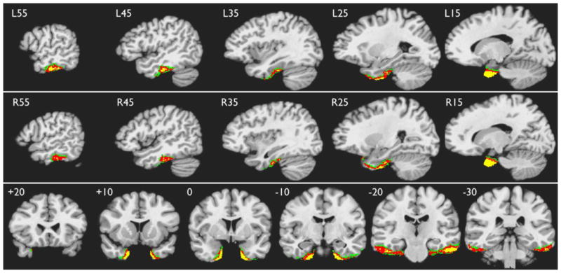
Removal of the anterior temporal lobe (ATL) is an effective surgical treatment for intractable temporal lobe epilepsy but carries a risk of language and verbal memory deficits. Preoperative localization of functional zones in the ATL might help reduce these risks, yet fMRI protocols in current widespread use produce very little activation in this region. Based on recent evidence suggesting a role for the ATL in semantic integration, we designed an fMRI protocol comparing comprehension of brief narratives (Story task) with a semantically shallow control task involving serial arithmetic (Math task). The Story > Math contrast elicited strong activation throughout the ATL, lateral temporal lobe, and medial temporal lobe bilaterally in an initial cohort of 18 healthy participants. The task protocol was then implemented at 6 other imaging centers using identical methods. Data from a second cohort of participants scanned at these centers closely replicated the results from the initial cohort. The Story-Math protocol provides a reliable method for activation of surgical regions of interest in the ATL. The bilateral activation supports previous claims that conceptual processing involves both temporal lobes. Used in combination with language lateralization measures, reliable ATL activation maps may be useful for predicting cognitive outcome in ATL surgery, though the validity of this approach needs to be established in a prospective surgical series.
Copyright © 2010 Elsevier Inc. All rights reserved.
Figures
References
-
- Baldo JV, Dronkers NF. Neural correlates of arithmetic and language comprehension: A common substrate? Neuropsychologia. 2007;45:229–235. - PubMed
-
- Baxendale S, Thompson P, Harkness W, Duncan J. Predicting memory decline following epilepsy surgery: A multivariate approach. Epilepsia. 2006;47:1887–1894. - PubMed
-
- Beeman M, Chiarello C, editors. Right hemisphere language comprehension: Perspectives from cognitive neuroscience. Erlbaum; Mahwah, NJ: 1998.
-
- Bell BD, Davies KG, Hermann BP, Walters G. Confrontation naming after anterior temporal lobectomy is related to age of acquisition of the object names. Neuropsychologia. 2000;38:83–92. - PubMed
Publication types
MeSH terms
Grants and funding
LinkOut - more resources
Full Text Sources
Other Literature Sources
