

Proliferation is a central independent prognostic factor and target for personalized and risk-adapted treatment in multiple myeloma
- PMID: 20884712
- PMCID: PMC3012769
- DOI: 10.3324/haematol.2010.030296
Proliferation is a central independent prognostic factor and target for personalized and risk-adapted treatment in multiple myeloma
Abstract
Background: Proliferation of malignant plasma cells is a strong adverse prognostic factor in multiple myeloma and simultaneously targetable by available (e.g. tubulin polymerase inhibitors) and upcoming (e.g. aurora kinase inhibitors) compounds.
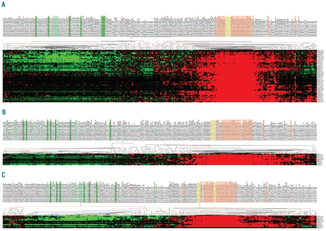
Design and methods: We assessed proliferation using gene expression-based indices in 757 samples including independent cohorts of 298 and 345 samples of CD138-purified myeloma cells from previously untreated patients undergoing high-dose chemotherapy, together with clinical prognostic factors, chromosomal aberrations, and gene expression-based high-risk scores.
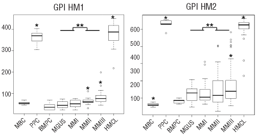
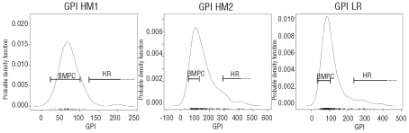
Results: In the two cohorts, 43.3% and 39.4% of the myeloma cell samples showed a proliferation index above the median plus three standard deviations of normal bone marrow plasma cells. Malignant plasma cells of patients in advanced stages or those harboring disease progression-associated gain of 1q21 or deletion of 13q14.3 showed significantly higher proliferation indices; patients with gain of chromosome 9, 15 or 19 (hyperdiploid samples) had significantly lower proliferation indices. Proliferation correlated with the presence of chromosomal aberrations in metaphase cytogenetics. It was significantly predictive for event-free and overall survival in both cohorts, allowed highly predictive risk stratification (e.g. event-free survival 12.7 versus 26.2 versus 40.6 months, P < 0.001) of patients, and was largely independent of clinical prognostic factors, e.g. serum β₂-microglobulin, International Staging System stage, associated high-risk chromosomal aberrations, e.g. translocation t(4;14), and gene expression-based high-risk scores.
Conclusions: Proliferation assessed by gene expression profiling, being independent of serum-β₂-microglobulin, International Staging System stage, t(4;14), and gene expression-based risk scores, is a central prognostic factor in multiple myeloma. Surrogating a biological targetable variable, gene expression-based assessment of proliferation allows selection of patients for risk-adapted anti-proliferative treatment on the background of conventional and gene expression-based risk factors.
Figures

References
-
- Kyle RA, Rajkumar SV. Multiple myeloma. N Engl J Med. 2004;351(18):1860–73. - PubMed
-
- Witzig TE, Timm M, Larson D, Therneau T, Greipp PR. Measurement of apoptosis and proliferation of bone marrow plasma cells in patients with plasma cell proliferative disorders. Br J Haematol. 1999;104 (1):131–7. - PubMed
-
- Boccadoro M, Gavarotti P, Fossati G, Pileri A, Marmont F, Neretto G, et al. Low plasma cell 3(H) thymidine incorporation in monoclonal gammopathy of undetermined significance (MGUS), smouldering myeloma and remission phase myeloma: a reliable indicator of patients not requiring therapy. Br J Haematol. 1984;58(4):689–96. - PubMed
-
- Greipp PR, Lust JA, O’Fallon WM, Katzmann JA, Witzig TE, Kyle RA. Plasma cell labeling index and beta 2-microglobulin predict survival independent of thymidine kinase and C-reactive protein in multiple myeloma. Blood. 1993;81(12):3382–7. - PubMed
-
- Greipp PR, Katzmann JA, O’Fallon WM, Kyle RA. Value of beta 2-microglobulin level and plasma cell labeling indices as prognostic factors in patients with newly diagnosed myeloma. Blood. 1988;72(1):219–23. - PubMed
Publication types
MeSH terms
Substances
LinkOut - more resources
Full Text Sources
Other Literature Sources
Medical
