. 2010 Nov;104(5):2766-77.
doi: 10.1152/jn.00491.2010. Epub 2010 Sep 8.

Endocannabinoid-dependent LTD in a nociceptive synapse requires activation of a presynaptic TRPV-like receptor

Affiliations

Endocannabinoid-dependent LTD in a nociceptive synapse requires activation of a presynaptic TRPV-like receptor

Sharleen Yuan et al. J Neurophysiol. 2010 Nov.

Abstract

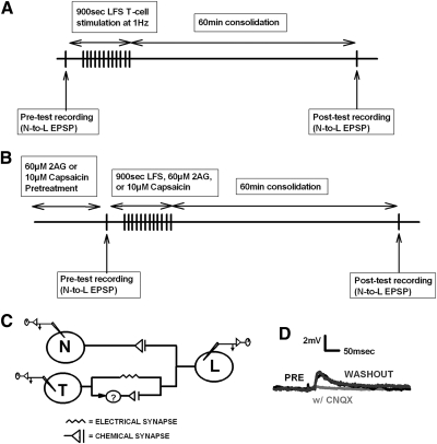
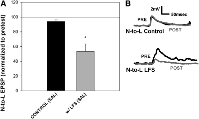
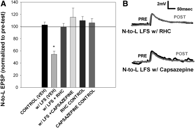
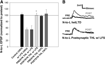
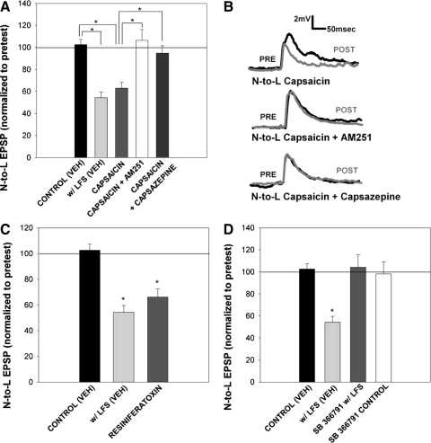
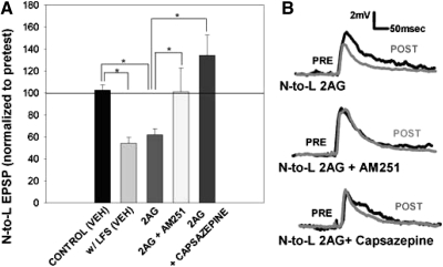
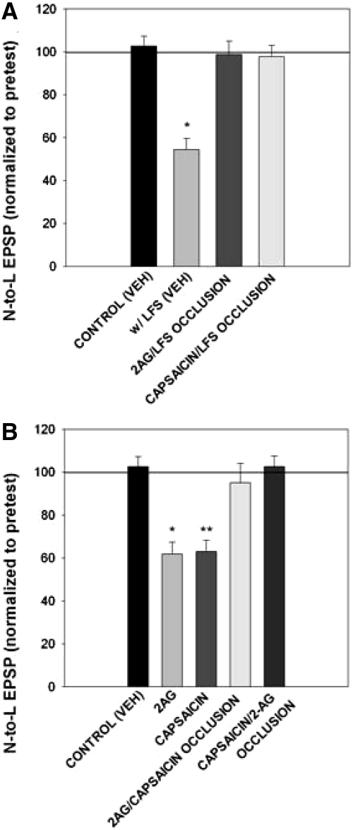
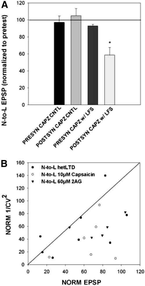
Recent studies have found that some forms of endocannabinoid-dependent synaptic plasticity in the hippocampus are mediated through activation of transient potential receptor vanilloid (TRPV) receptors instead of cannabinoid receptors CB1 or CB2. The potential role for synaptic localization of TRPV receptors during endocannabinoid modulation of nociceptive synapses was examined in the leech CNS where it is possible to record from the same pair of neurons from one preparation to the next. Long-term depression (LTD) in the monosynaptic connection between the nociceptive (N) sensory neuron and the longitudinal (L) motor neuron was found to be endocannabinoid-dependent given that this depression was blocked by RHC-80267, an inhibitor of DAG lipase that is required for 2-arachidonoyl glycerol (2AG) synthesis. Intracellular injection of a second DAG lipase inhibitor, tetrahyrdolipstatin (THL) was also able to block this endocannabinoid-dependent LTD (ecLTD) when injected postsynaptically but not presynaptically. N-to-L ecLTD was also inhibited by the TRPV1 antagonists capsazepine and SB 366791. Bath application of 2AG or the TRPV1 agonists capsaicin and resiniferatoxin mimicked LTD and both capsaicin- and 2AG-induced depression were blocked by capsazepine. In addition, pretreatment with 2AG or capsaicin occluded subsequent expression of LTD induced by repetitive activity. Presynaptic, but not postsynaptic, intracellular injection of capsazepine blocked both activity- and 2AG-induced ecLTD, suggesting that a presynaptic TRPV-like receptor in the leech mediated this form of synaptic plasticity. These findings potentially extend the role ecLTD to nociceptive synapses and suggest that invertebrate synapses, which are thought to lack CB1/CB2 receptor orthologues, utilize a TRPV-like protein as an endocannabinoid receptor.


Figures

Fig. 1.
Fig. 1.
Experimental protocols and synaptic circuitry. A: a pretest recording of the N-to-L or T-to-L synapse was made prior to low-frequency stimulation (LFS; 900 s, 1 Hz) of the T cell [or N cell if homosynaptic long-term depression (LTD) was being elicited]. In some experiments, the LFS was replaced by superfusion of 2-arachidonoyl glycerol (2AG), capsaicin, or resiniferatoxin for 900 s. Following a 60 min consolidation period, a posttest recording of the N-to-L or T-to-L synapse was carried out. B: during occlusion experiments, pretreatment with either 60 μM 2AG or 10 μM capsaicin was carried out prior to the pretest recordings of the N-to-L synapse. This was followed by LFS, 2AG (60 μM), or capsaicin (10 μM), depending on the experiment. Following a 60 min consolidation period, the posttest recordings of the N-to-L synapse were carried out. C: the nociceptive (N cell) sensory neuron has a monosynaptic chemical connection onto the longitudinal (L) motor neuron. The touch (T cell) sensory neuron has a monosynaptic electrical synapse and a polysynaptic chemical connection onto the L motor neuron; the interneuron(s) mediating the polysynaptic connection is unknown (?). D: 6-cyano-7-nitroquinoxalene-2,3-dione (CNQX) abolished the N-to-L excitatory postsynaptic potential (EPSP), indicating a glutamatergic synapse. Representative traces of the N-to-L synapse in normal saline (black line), during application of 20 μM of CNQX (gray line) and after 30 min washout in normal saline (dark gray line). Similar results were observed in CNQX experiments performed on the T-to-L synapse (data not shown) indicating that the polysynaptic chemical component of this circuit is glutamatergic.
Fig. 2.
Fig. 2.
Heterosynaptic LTD at the N-to-L synapse. A: bar graph showing that 15 min of LFS (1 Hz) of the T cell synapse produced a heterosynaptic LTD in the N-to-L synapse (n = 6) compared with controls tested without LFS (n = 9). Data were analyzed using a 1-way ANOVA [F(1,13) = 10.18; P < 0.01] with a post hoc Newman-Keuls test (*P < 0.01). B: sample traces showing no change in N-to-L EPSP amplitude in the control experiments (top) and a decrease in EPSP amplitude in synapses following T cell LFS (bottom). Pretest traces are indicated by a black line and posttest traces are represented by a gray line.
Fig. 3.
Fig. 3.
Heterosynaptic LTD is mediated by endocannabinoids and a transient potential receptor vanilloid (TRPV)-like receptor. A: bar graph showing the inhibition of heterosynaptic LTD through application of RHC-80267, a 2AG synthesis blocker, and capsazepine, a selective antagonist of TRPV1 receptors. Data were analyzed through a 1-way ANOVA [F(5,20) = 13.39; P < 0.01]. Bath application of 60 μM RHC (n = 4) or 10 μM capsazepine (n = 4) significantly blocked the heterosynaptic LTD observed with vehicle LFS [w/LFS (VEH)]. Post hoc Newman-Keuls tests detected a significant difference between veh LFS vs. veh control (*P < 0.05), vehicle LFS vs. LFS with RHC (*P < 0.05), vehicle LFS vs. LFS with capsazepine (*P < 0.05). There was no change in EPSP amplitude with RHC application alone (n = 3) or capsazepine application alone (n = 3), indicating that the drug itself did not have an effect. B, top: traces of the N-to-L synapse with RHC during LFS. Pretest traces (black line) show no changes compared with posttest traces (gray line) taken after stimulation. Bottom: traces of the N-to-L synapse with capsazepine during LFS. Pretest traces (black line) show no changes compared with posttest traces (gray line), indicating that capsazepine blocked heterosynaptic LTD.
Fig. 4.
Fig. 4.
LFS-induced LTD is mediated by postsynaptic endocannabinoid synthesis. A: bar graph showing tetrahyrdolipstatin (THL) iontophoresis in the pre- or postsynaptic neuron. Data were analyzed through a 1-way ANOVA [F(3,21) = 7.09; P < 0.01]. Newman-Keuls post hoc analyses indicated that iontophoretic injection of 10 μM THL into the postsynaptic L motor neuron (POSTSYN THL w/ LFS; n = 5) significantly blocked LFS-induced LTD compared with both presynaptic THL injection (PRESYN THL w/ LFS; n = 5; *P < 0.05) and LFS-induced LTD in saline [w/ LFS (VEH); *P < 0.05]. B, top: traces of the N-to-L synapse in saline during LFS. Posttest traces (gray lines) showing a significant depression in EPSP amplitude after LFS in saline compared with pretest traces (black lines). Bottom: traces of the N-to-L synapse after postsynaptic THL injection and LFS. Posttest traces (gray lines) show no change from pretest traces (black lines) after THL iontophoresis, even with LFS.
Fig. 5.
Fig. 5.
Capsaicin and resiniferatoxin mimicked LFS-induced LTD and was blocked by inhibitors of endocannabinoid and TRPV1 receptors. A: bar graph comparing the effects on synaptic transmission following treatment with either vehicle [control (VEH)], LFS in vehicle [w/LFS (VEH)], capsaicin alone (capsaicin, n = 12), capsaicin with 10 μM AM251 (capsaicin + AM251, n = 4), and capsaicin with 10 μM capsazepine (capsaicin + capsazepine, n = 5). One-way ANOVA [F(4,29) = 13.16; P < 0.01] detected a significant effect of treatment group. Newman-Keuls post hoc analysis detected a significant difference between the control (VEH) and w/LFS (VEH) groups (P < 0.05*) and between the capsaicin and control (VEH; P < 0.05*), capsaicin + AM251 (P < 0.05*), and capsaicin + capsazepine (P < 0.05*) groups. B, top: traces of the N-to-L synapse in the presence of 10 μM capsaicin. Baseline pretest traces (black line) show the EPSP amplitude before capsaicin application, while posttest traces (gray line) show a EPSP amplitude after treatment. Middle: traces of the N-to-L synapse in the presence of 10 μM capsaicin with 10 μM AM251. Posttest EPSP traces show no change from pretest EPSP traces if AM251 is applied with capsaicin, indicating that AM251 binds to TRPV receptors. Bottom: traces of the N-to-L synapse in the presence of 10 μM capsaicin and 10 μM capsazepine. Similar to AM251 traces, posttest EPSP amplitude did not differ from pretest EPSP amplitude if capsazepine is applied with capsaicin. C: bar graph comparing the effects on synaptic transmission following treatment with either vehicle [control (VEH)], LFS in vehicle [w/LFS (VEH)], and resiniferatoxin (n = 5). One-way ANOVA detected a significant effect of the resiniferatoxin treatment [F(2,14) = 20.73; P < 0.01]. Post hoc analysis using Newman-Keuls detected a significant difference between the control (VEH) group and w/LFS (VEH) group (*P < 0.05). Post hoc analysis also detected a significant difference between the control (VEH) group and the resiniferatoxin group (*P < 0.05). D: bar graph representing the effect of SB 366791 treatment on heterosynaptic LTD with vehicle [control (VEH)], LFS in vehicle [w/LFS (VEH)], LFS in the presence of SB 366791 (SB 366791 w/LFS; n = 5), and drug treatment control (SB 366791 control, n = 5). One-way ANOVA analysis indicated a significant main effect of the treatment group [F(3,18) = 9.68, P < 0.01]. Newman-Keuls post hoc analyses detected a significance difference between the vehicle LFS group with control (VEH; *P < 0.05) group and SB 366791 w/ LFS (*P < 0.05) group. Drug treatment alone (SB 366791 control) did not have any effect.
Fig. 6.
Fig. 6.
2AG mimicked LFS-induced LTD and was blocked by inhibitors of endocannabinoid and TRPV1 receptors. A: bar graph comparing the effects on synaptic transmission following treatment with either vehicle [control (VEH)], LFS in vehicle [w/LFS (VEH)], 2AG alone (2AG, n = 7), 2AG with 10 μM AM251 (2AG + AM251, n = 5), and 2AG with 10 μM capsazepine (2AG + capsazepine, n = 7). One-way ANOVA detected a significant effect of treatment group [F(4,24) = 8.08; P < 0.01]. Newman-Keuls post hoc analysis detected a significant difference between the w/LFS (VEH) group and the control (VEH; P < 0.05*) group and between the 2AG group and the control (VEH; P < 0.05*), 2AG + AM251 (P < 0.05*) and 2AG + capsazepine (P < 0.05*) groups. B, top: traces of the N-to-L synapse in the presence of 60 μM 2AG. Baseline pretest traces (black line) show the EPSP amplitude before 2AG application, while posttest traces (gray line) show EPSP amplitude after treatment. Middle: traces of the N-to-L synapse in the presence of 60 μM 2AG with 10 μM AM251. Posttest EPSP traces show no change from pretest EPSP traces if AM251 is applied with 2AG. Bottom: traces of the N-to-L synapse in the presence of 60 μM 2AG and 10 μM capsazepine. Similar to AM251 traces, posttest EPSP amplitude did not differ from pretest EPSP amplitude if capsazepine is applied with 2AG.
Fig. 7.
Fig. 7.
Pretreatment with 2AG or capsaicin occluded further depression. A: bar graph representing the effects of synaptic transmission with and without LFS [control (VEH); w/LFS (VEH)] compared with occlusions with 2AG pretreatment (2AG/LFS occlusion, n = 7) and capsaicin pretreatment (capsaicin/LFS occlusion, n = 5). Data were analyzed through a 1-way ANOVA [F(3,20) = 18.16; P < 0.01] for main treatment effect. Control groups had no change between pre- and posttest values, while LFS groups had a decrease in EPSP posttest amplitude. Pre- and posttest normalized values in the 2AG/LFS (n = 7) and capsaicin/LFS (n = 5) occlusion groups did not have an overall change. Pretest values were depressed due to 2AG or capsaicin pretreatment and subsequent LFS did not induce further depression, resulting in no change in posttest values. Newman-Keuls post hoc analysis detected a significant difference between the w/LFS (VEH) group and the control (VEH; P < 0.05*) group and between the LFS (veh) group with the 2AG/LFS occlusion (P < 0.05*) group and capsaicin/LFS occlusion (P < 0.05*) group. B: bar graph comparing occlusion experiments in control groups, control (VEH), 2AG alone (2AG), capsaicin alone (capsaicin), 2AG pretreatment with subsequent capsaicin application (2AG/capsaicin occlusion, n = 4), or capsaicin pretreatment with subsequent 2AG application (capsaicin/2AG occlusion, n = 5). Data were analyzed through a one-way ANOVA [F(4,29) = 11.99; P < 0.01] for main treatment effect. Control groups had no change between pretest and posttest values, while 2AG and capsaicin groups had a decrease in EPSP posttest amplitude. Pre- and posttest normalized values in the 2AG/capsaicin and capsaicin/2AG occlusion groups did not have an overall change leading to no change in normalized values. In the occlusion groups, pretest EPSPs were depressed after capsaicin or 2AG pretreatment and subsequent drug applications did not induce further depression, resulting in no change in posttest values. Newman-Keuls posthoc analysis detected a significant difference between the control (VEH) and 2AG (P < 0.05*) group and between the 2AG group with the 2AG/capsaicin occlusion (P < 0.05*) group and capsaicin/2AG occlusion (P < 0.05*) group. There was also a significant difference between the control (VEH) group and the capsaicin alone (P < 0.05**) groups and between the capsaicin group with the 2AG/capsaicin occlusion (P < 0.05**) group and capsaicin/2AG occlusion (P < 0.05**) group.
Fig. 8.
Fig. 8.
TPRV-like receptor-mediated endocannabinoid-dependent LTD (ecLTD) has a presynaptic locus. A: bar graph representing iontophoresis of 500 μM capsazepine pre- and postsynaptically without LFS (presyn capz cntl, n = 5; postsyn capz cntl, n = 3) and with LFS (presyn capz w/ LFS, n = 5; postsyn capz cntl, n = 8). One-way ANOVA revealed a main effect between the groups [F(3,17) = 7.23; P < 0.01]. Newman-Keuls post hoc analysis detected a significant difference between postsynaptic capsazepine iontophoresis with LFS with the controls (P < 0.05*) and presynaptic capsazepine iontophoresis with LFS (P < 0.05*). B: the normalized inverse CV2 was plotted in the y axis in relation to normalized EPSP amplitude in the x axis. N-to-L heterosynaptic LTD (●) mainly fell below the regression line, although 2 data points fell above the regression. Coefficient of variation analyses of capsaicin (○)- or 2AG (▾)-induced depression also fell below the regression line, indicating presynaptic expression.

Similar articles

Cited by

References

    1. Adams MD, Celniker SE, Holt RA, Evans CA, Gocayne JD, Amanatides PG, Scherer SE, Li PW, Hoskins RA, Galle RF, George RA, Lewis SE, Richards S, Ashburner M, Henderson SN, Sutton GG, Wortman JR, Yandell MD, Zhang Q, Chen LX, Brandon RC, Rogers YH, Blazej RG, Champe M, Pfeiffer BD, Wan KH, Doyle C, Baxter EG, Helt G, Nelson CR, Gabor GL, Abril JF, Agbayani A, An HJ, Andrews-Pfannkoch C, Baldwin D, Ballew RM, Basu A, Baxendale J, Bayraktaroglu L, Beasley EM, Beeson KY, Benos PV, Berman BP, Bhandari D, Bolshakov S, Borkova D, Botchan MR, Bouck J, Brokstein P, Brottier P, Burtis KC, Busam DA, Butler H, Cadieu E, Center A, Chandra I, Cherry JM, Cawley S, Dahlke C, Davenport LB, Davies P, de Pablos B, Delcher A, Deng Z, Mays AD, Dew I, Dietz SM, Dodson K, Doup LE, Downes M, Dugan-Rocha S, Dunkov BC, Dunn P, Durbin KJ, Evangelista CC, Ferraz C, Ferriera S, Fleischmann W, Fosler C, Gabrielian AE, Garg NS, Gelbart WM, Glasser K, Glodek A, Gong F, Gorrell JH, Gu Z, Guan P, Harris M, Harris NL, Harvey D, Heiman TJ, Hernandez JR, Houck J, Hostin D, Houston KA, Howland TJ, Wei MH, Ibegwam C, Jalali M, Kalush F, Karpen GH, Ke Z, Kennison JA, Ketchum KA, Kimmel BE, Kodira CD, Kraft C, Kravitz S, Kulp D, Lai Z, Lasko P, Lei Y, Levitsky AA, Li J, Li Z, Liang Y, Lin X, Liu X, Mattei B, McIntosh TC, McLeod MP, McPherson D, Merkulov G, Milshina NV, Mobarry C, Morris J, Moshrefi A, Mount SM, Moy M, Murphy B, Murphy L, Muzny DM, Nelson DL, Nelson DR, Nelson KA, Nixon K, Nusskern DR, Pacleb JM, Palazzolo M, Pittman GS, Pan S, Pollard J, Puri V, Reese MG, Reinert K, Remington K, Saunders RD, Scheeler F, Shen H, Shue BC, Siden-Kiamos I, Simpson M, Skupski MP, Smith T, Spier E, Spradling AC, Stapleton M, Strong R, Sun E, Svirskas R, Tector C, Turner R, Venter E, Wang AH, Wang X, Wang ZY, Wassarman DA, Weinstock GM, Weissenbach J, Williams SM, Woodage T, Worley KC, Wu D, Yang S, Yao QA, Ye J, Yeh RF, Zaveri JS, Zhan M, Zhang G, Zhao Q, Zheng L, Zheng XH, Zhong FN, Zhong W, Zhou X, Zhu S, Zhu X, Smith HO, Gibbs RA, Myers EW, Rubin GM, Venter JC. The genome sequence of Drosophila melanogaster. Science 287: 2185–2195, 2000 - PubMed
    1. Anwyl R. Induction and expression mechanisms of postsynaptic NMDA receptor-independent homosynaptic long-term depression. Prog Neurobiol 78: 17–37, 2006 - PubMed
    1. Bevan J, Hothi S, Hughes G, James IF, Rang HP, Shah K, Walpole CSJ, Yeats JC. Capsazepine: a competitive antagonist of the sensory neurone excitant capsaicin. Br J Pharmacol 107: 544–552, 1992 - PMC - PubMed
    1. Bisogno T, Howell F, Williams G, Minassi A, Cascio MG, Ligresti A, Matias I, Schiano-Moriello A, Paul P, Williams EJ, Gangadharan U, Hobbs C, Di Marzo V, Doherty P. Cloning of the first sn1-DAG lipases points to the spatial and temporal regulation of endocannabinoid signaling in the brain. J Cell Biol 163: 463–468, 2003 - PMC - PubMed
    1. Brockie PJ, Mellem JE, Hills T, Madsen DM, Maricq AV. The C. elegans glutamate receptor subunit NMR-1 is required for slow NMDA-activated currents that regulate reversal frequency during locomotion. Neuron 31: 617–630, 2001 - PubMed

Publication types

MeSH terms

LinkOut - more resources