Comparative Study
. 2010 Oct;85(10):881-97.
doi: 10.4065/mcp.2010.0326.

Autoimmune dementia: clinical course and predictors of immunotherapy response

Affiliations
Comparative Study

Autoimmune dementia: clinical course and predictors of immunotherapy response

Eoin P Flanagan et al. Mayo Clin Proc. 2010 Oct.

Abstract

Objective: To define the diagnostic characteristics and predictors of treatment response in patients with suspected autoimmune dementia.

Patients and methods: Between January 1, 2002, and January 1, 2009, 72 consecutive patients received immunotherapy for suspected autoimmune dementia. Their baseline clinical, radiologic, and serologic characteristics were reviewed and compared between patients who were responsive to immunotherapy and those who were not. Patients were classified as responders if the treating physician had reported improvement after immunotherapy (documented in 80% by the Kokmen Short Test of Mental Status, neuropsychological testing, or both).

Results: Initial immunotherapeutic regimens included methylprednisolone in 56 patients (78%), prednisone in 12 patients (17%), dexamethasone in 2 patients (3%), intravenous immune globulin in 1 patient (1%), and plasma exchange in 1 patient (1%). Forty-six patients (64%) improved, most in the first week of treatment. Thirty-five percent of these immunotherapy responders were initially diagnosed as having a neurodegenerative or prion disorder. Pretreatment and posttreatment neuropsychological score comparisons revealed improvement in almost all cognitive domains, most notably learning and memory. Radiologic or electroencephalographic improvements were reported in 22 (56%) of 39 patients. Immunotherapy responsiveness was predicted by a subacute onset (P<.001), fluctuating course (P<.001), tremor (P=.007), shorter delay to treatment (P=.005), seropositivity for a cation channel complex autoantibody (P=.01; neuronal voltage-gated potassium channel more than calcium channel or neuronal acetylcholine receptor), and elevated cerebrospinal fluid protein (>100 mg/dL) or pleocytosis (P=.02). Of 26 immunotherapy-responsive patients followed up for more than 1 year, 20 (77%) relapsed after discontinuing immunotherapy.

Conclusion: Identification of clinical and serologic clues to an autoimmune dementia allows early initiation of immunotherapy, and maintenance if needed, thus favoring an optimal outcome.


Figures

FIGURE 1.
FIGURE 1.
Improvements in Kokmen Short Test of Mental Status (STMS) score among responders after immunotherapy. Kokmen STMS scores improved in 32 of 46 patients responding to immunotherapy. Scores of nonresponders, by definition, did not improve. *Mild postimmunotherapy improvements in Kokmen STMS score were accompanied by significant improvements on neuropsychological testing.
FIGURE 2.
FIGURE 2.
Neuropsychological evaluations before and after treatment in 6 patients positive for voltage-gated potassium channel (VGKC) complex antibody. Mayo Older Americans Normative Studies (MOANS) scaled scores in 6 patients with VGKC complex antibodies illustrate the initial severity of neuropsychological impairment and posttreatment improvement. Patient 1 (patient 42 in Appendix 1, which appears at the end of this article) presented with severely impaired verbal memory and lexical fluency, intact verbal learning, and mild-to-moderate impairment of other indices; treatment was followed by substantial improvement in verbal memory and less impressive improvement in other scores. Patient 2 (patient 5 in Appendix 1) had severe impairment of verbal learning, verbal memory, and semantic fluency and showed a stepwise, protracted, but nevertheless complete recovery over 8 months. Patient 3 (patient 45 in Appendix 1) had impaired verbal learning and memory, semantic fluency, and executive function; all deficits except that in semantic fluency resolved after treatment. Patient 4 (patient 46 in Appendix 1) presented with impairment in all cognitive domains. After initial treatment with intravenous methylprednisolone, followed by intravenous immune globulin, the patient had mild initial improvement but marked cognitive fluctuation during the next 9 months; follow-up tests showed improvement in some areas and deterioration in others. Patients 5 (patient 33 in Appendix 1) and 6 (patient 35 in Appendix 1) both had dramatic clinical improvement after receiving intravenous corticosteroids but relapsed within 1 month. Cognitive testing during relapse showed impaired perceptual organization (PO) and verbal learning and memory, which resolved after resumption of treatment (completely in patient 5, incompletely in patient 6). Median VGKC complex autoantibody values for these 6 patients decreased significantly from 1.04 nmol/L (range, 0.13-4.22 nmol/L; reference range, 0.00-0.02 nmol/L) to 0.14 nmol/L (range, 0.00-1.87 nmol/L) after immunotherapy. CFT = Category Fluency Test; COWAT = Controlled Oral Word Association Test; DR = delayed recall; LOT = learning over trials; TMT = Trail-Making Test; VC = verbal comprehension.
FIGURE 3.
FIGURE 3.
Neuroimaging in patients with an immunotherapy-responsive cognitive disorder. Magnetic resonance imaging: yellow arrows indicate areas of abnormality on fluid-attenuated inversion recovery (FLAIR). A, 36-year-old woman (patient 10 in Appendix 1) had fluctuating memory problems and was seropositive for glutamic acid decarboxylase-65 autoantibody. Bilateral hippocampal axial FLAIR abnormality, shown in A1, almost completely resolved after treatment with intravenous (IV) methylprednisolone (A2). B, 51-year-old woman (patient 20 in Appendix 1) had subacute fluctuating memory problems, multifocal neurologic examination findings, and evidence of autoimmunity (IgM antiphospholipid antibody). Symmetric confluent T2 signal abnormality in the white matter of both hemispheres (B1) decreased after treatment with IV methylprednisolone (B2). C, 60-year-old man (patient 41 in Appendix 1) had memory, language, and gait problems and was seropositive for both striational and glutamic acid decarboxylase-65 antibodies. Axial T1 magnetic resonance imaging with contrast demonstrated periventricular vessel enhancement (C1) and resolution after treatment (C2). D, 53-year-old woman (patient 29 in Appendix 1) had memory loss, hallucinations, and subsequent seizure; cerebrospinal fluid protein was elevated (>100 mg/dL), and she was seropositive for thyroid peroxidase antibodies and neuronal and muscle acetylcholine receptor antibodies. Axial FLAIR images show diffusely increased T2 signal in the midbrain (D1), which improved after treatment with IV methylprednisolone (D2). Multiple myeloma was diagnosed 18 months after neurologic presentation. Positron emission tomographic imaging: Brain reconstructions (brighter color represents regions of hypometabolism) in a 58-year-old man (patient 21 in Appendix 1) who presented with personality change and memory problems and had elevated cerebrospinal fluid protein (>100 mg/dL). Hypometabolism, predominantly frontal and temporal (E1), improved after treatment with IV methylprednisolone (E2). Single-photon emission computed tomographic brain imaging: Brain neuroimaging in a 35-year-old man (patient 22 in Appendix 1) who presented with vertigo and memory problems, had multiple coexisting autoimmune conditions, and was seropositive for muscle acetylcholine receptor and striational antibodies. Diffuse decrease in uptake in frontotemporoparietal regions (F1) was markedly improved globally after treatment with IV methylprednisolone (F2).
FIGURE 4.
FIGURE 4.
Electroencephalogram (EEG) before and after immunotherapy in patients with immunotherapy-responsive cognitive disorders. A, EEG in a 60-year-old man (patient 31 in Appendix 1) who presented with memory difficulty and inattention and had elevated cerebrospinal fluid protein (>100 mg/dL) and voltage-gated potassium channel complex autoantibody. Before treatment, the EEG showed a left anterior temporal lobe seizure (A1) and left temporal intermittent rhythmic delta and sharp wave activity (A2). This resolved after treatment with both a loading dose of phenytoin and intravenous methylprednisolone (A3). Of note, this patient had corresponding improvements in mesiotemporal signal abnormality on magnetic resonance imaging and resolution of left temporal hypermetabolism on positron emission tomography. B, EEG in a 71-year-old man (patient 13 in Appendix 1) who presented with subacute, rapidly progressing memory problems mimicking Creutzfeldt-Jakob disease. He was seropositive for thyroid peroxidase antibody and had inflammatory cerebrospinal fluid. Note severe, diffuse theta and delta wave slowing maximal over the left hemisphere (B1), and improvement with mild background slowing (7-8 Hz posterior alpha) after treatment with intravenous methylprednisolone (B2).
FIGURE 5.
FIGURE 5.
IgGs binding selectively to central nervous system tissues. Immunohistochemical staining by glutamic acid decarboxylase-65 (A-C; patient 29 in Appendix 1 and patients 53 and 70 in Appendix 2) and voltage-gated potassium channel complex (D, E; patients 31 and 33 in Appendix 1) antibodies on a composite of mouse tissues: kidney (Kid), cerebellum (CbL, both granular layers [GL] and molecular layers [ML]), midbrain (MB), entorhinal cortex (Cortex), hippocampus (Hip), and thalamus (Thal). F, Unclassified IgG restricted to central nervous system tissues in a patient with immunotherapy-responsive dementia (patient 14 in Appendix 1). No patient had an IgG targeting N-methyl-d-aspartate, α-amino-3-hydroxy-5-methyl-4-isoxazolepropionic acid, or γ-aminobutyric type B receptors.
FIGURE 6.
FIGURE 6.
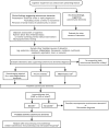
Diagnostic algorithm for suspected autoimmune dementia or encephalopathy. CSF = cerebrospinal fluid; IVIG = intravenous immune globulin; IVMP = intravenous methylprednisolone. * If no other cause for dementia is identified, consider a trial of immunotherapy. If findings on neural antibody testing are positive, consider screening for underlying malignancy with computed tomography or positron emission tomography. CSF protein level >100 mg/dL or pleocytosis (white blood cell count >5). § Reference . // Repeat objective cognitive assessments to aid in evaluating response to immunotherapy.

Comment in

  • Autoimmune encephalopathy.
    Caselli RJ, Drazkowski JF, Wingerchuk DM. Caselli RJ, et al. Mayo Clin Proc. 2010 Oct;85(10):878-80. doi: 10.4065/mcp.2010.0536. Mayo Clin Proc. 2010. PMID: 20884823 Free PMC article. No abstract available.

References

    1. McKeon A, Lennon VA, Pittock SJ. Immunotherapy-responsive dementias and encephalopathies. Continuum Lifelong Learning Neurol. 2010;16(2):80-101 - PubMed
    1. Castillo P, Woodruff B, Caselli R, et al. Steroid-responsive encephalopathy associated with autoimmune thyroiditis. Arch Neurol. 2006;63(2):197-202 - PubMed
    1. Geschwind MD, Tan KM, Lennon VA, et al. Voltage-gated potassium channel autoimmunity mimicking Creutzfeldt-Jakob disease. Arch Neurol. 2008;65(10):1341-1346 - PMC - PubMed
    1. McKeon A, Marnane M, O'Connell M, Stack JP, Kelly PJ, Lynch T. Potassium channel antibody associated encephalopathy presenting with a frontotemporal dementia like syndrome. Arch Neurol. 2007;64(10):1528-1530 - PubMed
    1. Brain L, Jellinek EH, Ball K. Hashimoto's disease and encephalopathy. Lancet. 1966;2(7462):512-514 - PubMed

Publication types