Review
. 2010 Oct;126(4):1172-1180.
doi: 10.1097/PRS.0b013e3181eae781.

Scarless fetal wound healing: a basic science review

Affiliations
Review

Scarless fetal wound healing: a basic science review

Barrett J Larson et al. Plast Reconstr Surg. 2010 Oct.

Abstract

Scar formation is a major medical problem that can have devastating consequences for patients. The adverse physiological and psychological effects of scars are broad, and there are currently no reliable treatments to prevent scarring. In contrast to adult wounds, early gestation fetal skin wounds repair rapidly and in the absence of scar formation. Despite extensive investigation, the exact mechanisms of scarless fetal wound healing remain largely unknown. For some time, it has been known that significant differences exist among the extracellular matrix, inflammatory response, cellular mediators, and gene expression profiles of fetal and postnatal wounds. These differences may have important implications in scarless wound repair.


Conflict of interest statement

Disclosure: The authors have no commercial associations or financial disclosures that might pose or create a conflict of interest with information presented in this article.

Figures

Fig. 1
Fig. 1
Histology of human fetal skin at 14 and 20 weeks of gestation and adult skin. (Left) At 14 weeks, the epidermis consists of a basal layer, an intermediate cell layer, and a periderm. (Center) At 20 weeks, the number of intermediate cell layers has increased, and developing hair follicles are visible. (Right) Adult skin has a less cellular dermis and a multilayered epidermis with basal, spinous, granular, and cornified layers. Scale bars = 100 μm. Reproduced with permission from Springer from Coolen NA, Schouten KC, Middelkoop E, et al. Comparison between human fetal and adult skin. Arch Dermatol Res. 2010;302:47–55.
Fig. 2
Fig. 2
Scarless healing of E16 fetal mouse wounds (hematoxylin and eosin stain). Black arrows indicate the India ink tattoo made at the time of wounding to demonstrate scarless wound location. Healed wounds (above, left and below, left) at 72 hours (100×). The epidermal appendage (developing hair follicles) pattern shows numerous appendages directly in the healed wound. Magnified views of the same wounds (above, right and below, right) showing epidermal appendages (open arrows) within the wound site (200×). No inflammatory infiltrate is present. Reproduced from Beanes SR, Hu FY, Soo C, et al. Confocal microscopic analysis of scarless repair in the fetal rat: Defining the transition. Plast Reconstr Surg. 2002;109:160–170.
Fig. 3
Fig. 3
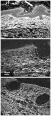
Scarless healing of E16 fetal rat wounds (confocal microscopy). Collagen fibers are stained with sirius red and appear white. (Above) Healed wound harvested at 72 hours (200×). The epidermis is thickened at the wound site (arrow). The collagen fiber is reticular and unchanged from the surrounding dermis. (Center) Healed wound harvested at 72 hours under a higher magnification (1000 ×). The collagen fibers are thin and closely approximating each other with little interfiber space. The fibers are arranged in a wispy reticular pattern. (Below) Non-wounded E19 skin at the same magnification as B (1000×). The dermal collagen fiber pattern is identical to the center image. Reproduced from Beanes SR, Hu FY, Soo C, et al. Confocal microscopic analysis of scarless repair in the fetal rat: Defining the transition. Plast Reconstr Surg. 2002;109:160–170.
Fig. 4
Fig. 4
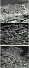
Early scar formation after the transition point in E18 fetal rat wounds (confocal microscopy). Collagen fibers are stained with sirius red and appear white. (Above) Healed wound at 72 hours (200×). The wound dermal collagen pattern (open triangles) is different from the surrounding nonwounded dermis (green arrow). The fibers are less densely compacted. No epidermal appendages are present. Neovascularization is shown with the white arrows. (Center) Healed wound at 72 hours at a higher magnification (1000×). The collagen fibers are thicker but with greater interfiber spaces compared with nonwounded dermis. (Below) Nonwounded skin at E21 days gestational age (1000 ×). When compared with wound collagen fibers (center), non-wounded dermal collagen fibers are thinner with less interfiber space. Reproduced from Beanes SR, Hu FY, Soo C, et al. Confocal microscopic analysis of scarless repair in the fetal rat: Defining the transition. Plast Reconstr Surg. 2002;109:160–170.

Similar articles

Cited by

References

    1. Colwell AS, Phan TT, Kong W, et al. Hypertrophic scar fibroblasts have increased connective tissue growth factor expression after transforming growth factor-beta stimulation. Plast Reconstr Surg. 2005;116:1387–1390. - PubMed
    1. Madden JW, Peacock EE. Studies on the biology of collagen during wound healing: Dynamic metabolism of scar collagen and remodeling of dermal wounds. Ann Surg. 1971;174:511–520. - PMC - PubMed
    1. Rowlatt U. Intrauterine healing in a 20-week human fetus. Virchows Arch. 1979;381:353–361. - PubMed
    1. Adzick NS, Longaker MT. Characteristics of Fetal Tissue Repair. New York: Elsevier; 1992.
    1. Lane AL. Human fetal skin development. Pediatr Dermatol. 1986;3:487–491. - PubMed