. 2011 Mar;18(3):493-505.
doi: 10.1038/cdd.2010.120. Epub 2010 Oct 1.

Novel role of KCNQ2/3 channels in regulating neuronal cell viability

Affiliations

Novel role of KCNQ2/3 channels in regulating neuronal cell viability

X Zhou et al. Cell Death Differ. 2011 Mar.

Abstract

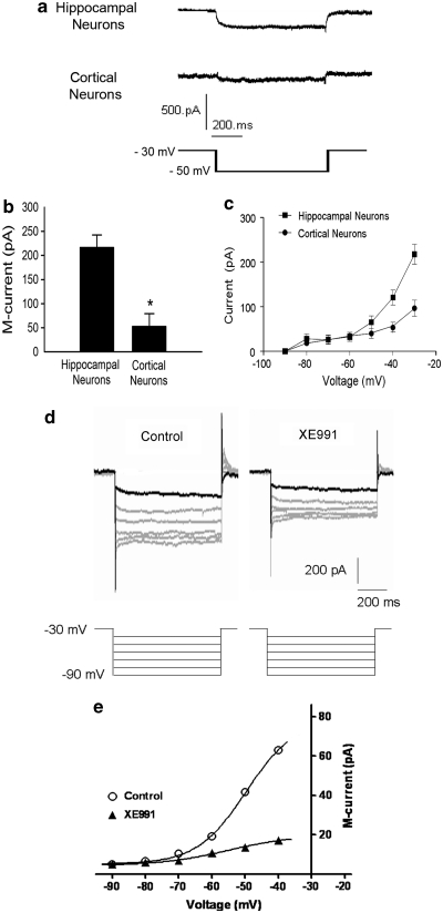
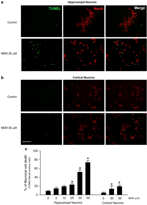
Overactivation of certain K(+) channels can mediate excessive K(+) efflux and intracellular K(+) depletion, which are early ionic events in apoptotic cascade. The present investigation examined a possible role of the KCNQ2/3 channel or M-channel (also named Kv7.2/7.3 channels) in the pro-apoptotic process. Whole-cell recordings detected much larger M-currents (212 ± 31 pA or 10.5 ± 1.5 pA/pF) in cultured hippocampal neurons than that in cultured cortical neurons (47 ± 21 pA or 2.4 ± 0.8 pA/pF). KCNQ2/3 channel openers N-ethylmaleimide (NEM) and flupirtine caused dose-dependent K(+) efflux, intracellular K(+) depletion, and cell death in hippocampal cultures, whereas little cell death was induced by NEM in cortical cultures. The NEM-induced cell death was antagonized by co-applied KCNQ channel inhibitor XE991 (10 μM), or by elevated extracellular K(+) concentration. Supporting a mediating role of KCNQ2/3 channels in apoptosis, expression of KCNQ2 or KCNQ2/3 channels in Chinese hamster ovary (CHO) cells initiated caspase-3 activation. Consistently, application of NEM (20 μM, 8 h) in hippocampal cultures similarly caused caspase-3 activation assessed by immunocytochemical staining and western blotting. NEM increased the expression of extracellular signal-regulated protein kinases 1 and 2 (ERK1/2), induced mitochondria membrane depolarization, cytochrome c release, formation of apoptosome complex, and apoptosis-inducing factor (AIF) translocation into nuclear. All these events were attenuated by blocking KCNQ2/3 channels. These findings provide novel evidence that KCNQ2/3 channels could be an important regulator in neuronal apoptosis.


Figures

Figure 1
Figure 1
KCNQ2 and KCNQ3 subunits in cultured hippocampal and cortical neurons. Expression and functional activity of KCNQ2 and KCNQ3 channels were tested in hippocampal and cortical neurons of 14 days in vitro (DIV). (a, b) Immunostaining for KCNQ2 (red), KCNQ3 (red), and Hoechst (blue) in hippocampal neurons (a) and cortical neurons (b). KCNQ2/3 was detected in both the cell body and dendrites of hippocampal and cortical neurons. Scale bar=10 μm. (c) Western blotting showed that there is no difference in KCNQ2/3 expression level between cortical (Cort) and hippocampal (Hipp) neurons. n=3 independent experiments. Error bars represent S.E.M.
Figure 2
Figure 2
M-currents in cultured hippocampal and cortical neurons. M-currents were recorded in 14–17 DIV hippocampal and cortical neurons using whole-cell recording. (a) Representative M-current traces recorded from hippocampal and cortical neurons, respectively. The recording of M-currents was started from a holding potential of −30 mV to allow the channels in activated state. A hyperpolarized pulse to −50 mV then close the M-channel, this hyperpolarization does not activate other voltage-gated channels that are sensitive only to depolarization. The M-current was also identified by its K+ dependence, sensitivity to block by muscarine and XE991, its slow activation phase and non-inactivation feature., (b) The bar graph shows mean values of M-currents in hippocampal neurons (n=10) and in cortical neurons (n=7). The current was enhanced by NEM in a dose-dependent manner, the enhanced current remained sensitive to block by XE991 (data not shown). (c) The I-V relationship of M-currents in hippocampal and cortical neurons. Although the amplitude of the currents was different in these two types of cells, both show typical voltage sensitivity and outward rectification features of the M-current. N=5 for each group. Error bars represent S.E.M. *P<0.05 versus hippocampal neurons. (d) M-currents (upper panel) in a hippocampal neuron triggered at different voltage steps (down panel) and their block by XE991 (5 μM). Current traces were from the same cell before and after 10 min application of XE991. (e) I-V curves of M-currents before and after 5 μM XE991. The I-V curves were constructed by plotting the amplitude of inward relaxation upon hyperpolarizing pulses. XE991 at 5 μM substantially (70–80%) blocked the current in 10 min. N=3 cells
Figure 3
Figure 3
KCNQ channel activation induced neuronal cell death in cultured hippocampal and cortical neurons. Cell viability in the presence of the KCNQ channel opener, NEM (30 or 50 μM, 24 h), was assessed by TUNEL staining. (a, b) NEM (30 μM) caused more TUNEL-positive cells in hippocampal neurons (a) than in cortical neurons (b). Scale bar=100 μm. (c) Quantification of the percentage of TUNEL-positive neurons in hippocampal and cortical cultures. NEM caused dose-dependent significant cell death in hippocampal cultures, whereas much less cell death was detected in cortical neurons. Mean±S.E.M. N≥3 independent experiments. *P<0.05 versus vehicle controls; #P<0.05, versus NEM (30 or 50 μM) treatment in hippocampal cells
Figure 4
Figure 4
Protective effects of KCNQ blocker or high external K+ on NEM and flupirtine-induced hippocampal neuronal death. (a) KCNQ channel inhibitor XE991 (10 μM) or high K+ external solution (25 mM KCl) showed protective effect on 30 μM NEM-induced cell death in hippocampal neurons, revealed using TUNEL assay. XE991 (10 μM) alone did not show significant effect on the basal level of cell viability. (b) KCNQ channel opener flupirtine (100 μM) cause more neuronal death compared with controls, and XE991 blocked flupirtine toxicity. Mean±S.E. N≥3 independent experiments. (c, d) PBFI-AM imaging was applied to measure intracellular K+ changes. There was no change in control cells (c) but significant decreases in PBFI fluorescence were detected after exposure to either NEM (30 μM, 80 min) (d) or the K+ ionophore valinomycin (50 μM, 80 min) (e). Arrows point to PBFI imaging in representative cells under control and NEM conditions. (e) The bar graph summarizes experiments in (c) (n=3 independent measurements in each group). *P<0.05 versus vehicle controls; #P<0.05 versus NEM treatment
Figure 5
Figure 5
Effects of expression of KCNQ2/KCNQ3 channels on caspase-3 activation. (ad) KCNQ2 and KCNQ3 subunits were subcloned into pEGFP-C3 (a) and pDsRed2-C1 vectors (c) and transfected into CHO cells shown as green and red color in transfected cells (b, d). (e) The expression of KCNQ2 alone or KCNQ2/3 induced activation of caspase-3, and the KCNQ channel blocker XE991 suppressed caspase-3 activation in KCNQ-transfected CHO cells. Expression of homogenous KCNQ3 showed much less effect on caspase-3 activation, which is in line with previous reports that this subunit alone may not form functional M-channels (see text for discussion). *P<0.05 versus control vector transfection; #P<0.05 versus KCNQ expression
Figure 6
Figure 6
KCNQ channel opener-induced increases in ERK phosphorylation and caspase-3 activation. The effects of NEM on apoptosis-related ERK phosphorylation and caspase-3 activation were detected in hippocampal cultures. (ad) Representative western blotting showed expression levels of phosphor-ERK1/2 (44/42 KD) and total ERK1/2 (44/42KD) (a, b) or cleaved caspase-3 (c, d) in control, NEM (20 μM), or NEM (20 μM) plus XE991 (10 μM)-treated hippocampal neurons. XE991 drastically prevented NEM-increased ERK1/2 phosphorylation and caspase-3 activation. Mean measurement of ERK1/2 intensities was quantified. Mean±S.E.M. N≥3 independent assays. (e) Immunostaining of caspase-3 (red) or NeuN (green) were shown in control, NEM (20 μM) or NEM plus XE991 (10 μM). (f) Quantification of capase-3-positive cells. XE991 significantly decreased NEM-induced caspase-3 activation. Scale bar=100 μm. *P<0.05 versus vehicle controls; #P<0.05 versus NEM
Figure 7
Figure 7
Inhibition of ERK activity reduced neuronal death and caspase-3 activation in hippocampal neurons. Neuronal cell death and caspase activation were tested in hippocampal neurons using TUNEL staining and western blot analysis. (a) Exposure to NEM (20 μM) for 24 h caused about 35% cell death in hippocampal cultures measured by TUNEL assay. Both XE991 (10 μM) and UO126 (10 μM) showed protective effects against NEM toxicity. (b, c) Western blotting using the antibody at cleaved caspase-3 showed increased caspase-3 activation in hippocampal neurons after 8 h exposure to 20 μM NEM event was attenuated by XE991 (10 μM) or UO126 (10 μM). Mean±S.E.M.; N≥3 independent assays. *P<0.05 versus vehicle controls; #P<0.05 versus NEM treatment
Figure 8
Figure 8
Effects of KCNQ channel opener on mitochondria membrane potential. The mitochondrial membrane potential (ΔΨ) was monitored using TMRM staining. Hippocampal neurons were preloaded with TMRM (200 nM) 30 min before NEM (20 μM) treatment at indicated times. The intensity of TMRM fluorescence was detected using a fluorescent microscope. (a) NEM induced a dramatic collapse of the mitochondria membrane potential within 30 min. This mitochondria membrane depolarization was significantly blocked by XE991 (10 μM). (b) NEM-induced mitochondria membrane depolarization was partially inhibited by XE991. Mean±S.E.M.; N≥3 separate experiments. *P<0.05 versus vehicle controls; #P<0.05 versus NEM treatment
Figure 9
Figure 9
Effects of KCNQ channel opener on cytosolic release and nuclear translocation of cytochrome c and AIF. Western blot analysis of cytochrome c and AIF in cytoslic and nuclear compartments after NEM exposure. (a, b) Hippocampal neurons were treated with NEM (20 μM) for 4 h. Cytosolic proteins (a) and nuclear proteins (b) were separated from mitochondria proteins and subjected to western blotting. (ce) Summary of (a, b) NEM-induced cytochrome c release from mitochondria was blocked by XE991 (10 μM) or UO126 (10 μM) (c). XE991 and UO126 also significantly attenuated nuclear translocation of cytochrome c induced by NEM (d). The nuclear translocation of AIF was also largely prevented by co-applied XE991 or UO126 (e). Mean±S.E.M.; N≥3 separate assays. *P<0.05 versus vehicle controls; #P<0.05 versus NEM treatment
Figure 10
Figure 10
KCNQ channel activation induced apoptosome formation in hippocampal neurons. Protein extractions from hippocampal neurons were collected and then used in immunoprecipitation assays against Apaf-1. Western blotting was then performed to detect the level of caspase-9 to indicate the formation of apoptosome complex. Note that the y axis value 1.0 is the basal level of apoptosome complex in control cells. Apoptosome formation was observed after NEM exposure, which was completely prevented by co-applied XE991 (10 μM). Mean±S.E.M.; N≥3 separate experiments; *P<0.05 versus vehicle controls; #P<0.05 versus NEM treatment

Similar articles

Cited by

References

    1. Burg ED, Remillard CV, Yuan JX. K+ channels in apoptosis. J Membr Biol. 2006;209:3–20. - PubMed
    1. Taatjes DJ, Sobel BE, Budd RC. Morphological and cytochemical determination of cell death by apoptosis. Histochem Cell Biol. 2008;129:33–43. - PMC - PubMed
    1. Yu SP. Regulation and critical role of potassium homeostasis in apoptosis. Prog Neurobiol. 2003;70:363–386. - PubMed
    1. Yu SP, Choi DW. Ions, cell volume, and apoptosis. Proc Natl Acad Sci USA. 2000;97:9360–9362. - PMC - PubMed
    1. Yu SP, Yeh CH, Sensi SL, Gwag BJ, Canzoniero LM, Farhangrazi ZS, et al. Mediation of neuronal apoptosis by enhancement of outward potassium current. Science. 1997;278:114–117. - PubMed

Publication types

MeSH terms