. 2011 Mar;18(3):479-92.
doi: 10.1038/cdd.2010.118. Epub 2010 Oct 1.

Regulation of autophagic activity by 14-3-3ζ proteins associated with class III phosphatidylinositol-3-kinase

Affiliations

Regulation of autophagic activity by 14-3-3ζ proteins associated with class III phosphatidylinositol-3-kinase

M Pozuelo-Rubio. Cell Death Differ. 2011 Mar.

Abstract

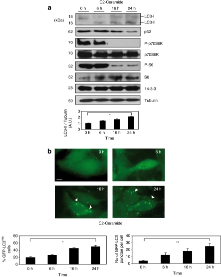
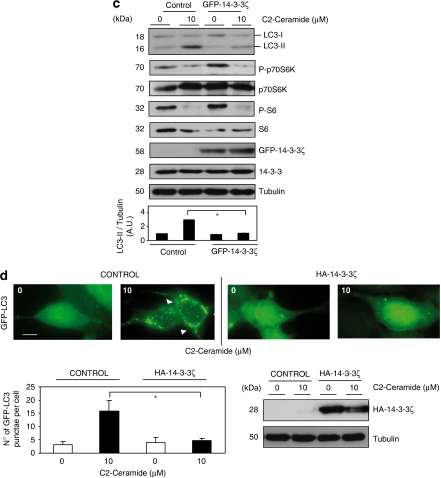
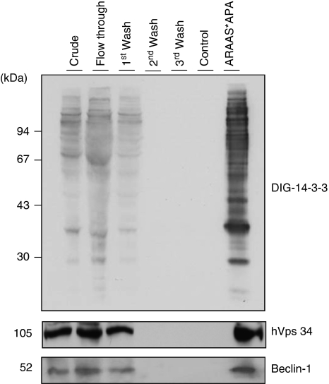
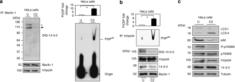
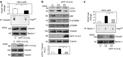
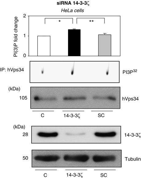
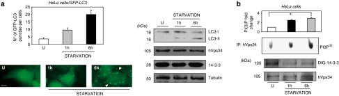
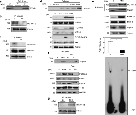
14-3-3s are binding proteins with survival functions in cells by interaction with proteins involved in the regulation of cell fate. The role of 14-3-3 during autophagy was investigated, thus, a forced expression of 14-3-3ζ reduces C2-ceramide-induced autophagy, whereas depletion of 14-3-3ζ promotes autophagy. The 14-3-3 role in autophagyc-related proteins was also investigated. The human vacuolar protein sorting 34 (hVps34), the class III phosphatidylinositol-3-kinase mediates multiple vesicle-trafficking processes such as endocytosis and autophagy, its activation being a requirement for autophagy initiation. Using chromatography techniques, hVps34 were eluted from a 14-3-3 affinity column, showing also a direct interaction with 14-3-3 proteins under physiological condition. Further analysis suggests that hVps34/14-3-3 association is a phorbol-12-myristate-13-acetate-dependent phosphorylated mechanism promoting a strong inhibition of the hVps34 lipid kinase activity, proteins kinase C being the likely kinase involved in phosphorylation and 14-3-3 binding of hVps34 under physiological conditions. Meanwhile, stimulation of autophagy leads to the dissociation of the 14-3-3/hVps34 complex enhancing hVps34 lipid kinase activity. Forced expression of 14-3-3ζ reduces hVps34 kinase activity and depletion of 14-3-3ζ promotes upregulation of this activity. In this study, 14-3-3ζ proteins are shown as a negative regulator of autophagy through regulation of a key component of early stages of the autophagy pathway, such as hVps34.


Figures

Figure 1
Figure 1
Overexpression of 14-3-3ζ proteins block C2-ceramide-induced autophagy. (a) Hela cells were incubated with 10 μM of C2-ceramide for indicated times. Cell extracts were harvested for immunoblot analysis of LC3-I processing into LC3-II, p62/SQSTM1, P-p70S6K, total p70S6K, P-S6, total 6S and 14-3-3 proteins. Tubulin was used as a protein loading control. Endogenous LC3-II/tubulin levels were quantified and the ratio presented as arbitrary units (N=3, *P=0.0006, Student t-test). (b) HEK293T cells transfected with GFP-LC3 were treated as in (a) and GFP-LC3 was analyzed by microscopy. Quantification of GFP-LC3vac cells number and number of GFP-LC3 punctae per cell were performed by counting in a blinded experiment. Columns, average of three different experiments, set up with duplicated samples and counting 6 × 100 cells per sample (quantification of GFP-LC3vac cells with more that five punctae per cell, is presented as a percentage of total cell numbers, N=3, *P=0.0001, **P=0.00087 Student t-test). (c) HEK293T cells transfected with GFP (control) or GFP-14-3-3ζ (GFP-14-3-3ζ) were left untreated or stimulated with C2-ceramide 10 μM for 24 h. Cell extracts were harvested for immunoblot analysis of LC3-I processing into LC3-II, P-p70S6K, total p70S6K, P-S6, total 6S, GFP-14-3-3ζ and total 14-3-3 proteins. Tubulin was used as a protein loading control. Endogenous LC3-II/tubulin levels were quantified and the ratio presented as arbitrary units. Columns, average of three different experiments (*P=0.00037, Student t-test). (d) HEK293T/GFP-LC3 cells were transfected with HA-control or HA-14-3-3ζ were left untreated or stimulated with C2-ceramide 10 μM for 24 h. GFP-LC3 was analyzed. Quantification of number of GFP-LC3 punctae per cell was performed by counting in a blinded experiment. Columns, average of three different experiments, set up with duplicated samples and counting 6 × 100 cells per sample, shown as percentage. Bars=10 μm (*P=0.00059, Student t-test). Western blot analysis shows levels of HA-14-3-3ζ. Tubulin was used as a loading control. A full-color version of this figure is available at Cell Death and Differentiation online
Figure 1
Figure 1
Overexpression of 14-3-3ζ proteins block C2-ceramide-induced autophagy. (a) Hela cells were incubated with 10 μM of C2-ceramide for indicated times. Cell extracts were harvested for immunoblot analysis of LC3-I processing into LC3-II, p62/SQSTM1, P-p70S6K, total p70S6K, P-S6, total 6S and 14-3-3 proteins. Tubulin was used as a protein loading control. Endogenous LC3-II/tubulin levels were quantified and the ratio presented as arbitrary units (N=3, *P=0.0006, Student t-test). (b) HEK293T cells transfected with GFP-LC3 were treated as in (a) and GFP-LC3 was analyzed by microscopy. Quantification of GFP-LC3vac cells number and number of GFP-LC3 punctae per cell were performed by counting in a blinded experiment. Columns, average of three different experiments, set up with duplicated samples and counting 6 × 100 cells per sample (quantification of GFP-LC3vac cells with more that five punctae per cell, is presented as a percentage of total cell numbers, N=3, *P=0.0001, **P=0.00087 Student t-test). (c) HEK293T cells transfected with GFP (control) or GFP-14-3-3ζ (GFP-14-3-3ζ) were left untreated or stimulated with C2-ceramide 10 μM for 24 h. Cell extracts were harvested for immunoblot analysis of LC3-I processing into LC3-II, P-p70S6K, total p70S6K, P-S6, total 6S, GFP-14-3-3ζ and total 14-3-3 proteins. Tubulin was used as a protein loading control. Endogenous LC3-II/tubulin levels were quantified and the ratio presented as arbitrary units. Columns, average of three different experiments (*P=0.00037, Student t-test). (d) HEK293T/GFP-LC3 cells were transfected with HA-control or HA-14-3-3ζ were left untreated or stimulated with C2-ceramide 10 μM for 24 h. GFP-LC3 was analyzed. Quantification of number of GFP-LC3 punctae per cell was performed by counting in a blinded experiment. Columns, average of three different experiments, set up with duplicated samples and counting 6 × 100 cells per sample, shown as percentage. Bars=10 μm (*P=0.00059, Student t-test). Western blot analysis shows levels of HA-14-3-3ζ. Tubulin was used as a loading control. A full-color version of this figure is available at Cell Death and Differentiation online
Figure 2
Figure 2
Downregulation of endogenous 14-3-3ζ sensitizes cells to autophagy. (a) HeLa cells expressing GFP-LC3 were transfected either with siRNA oligonucleotide targeting 14-3-3ζ or with a scrambled RNA oligonucleotide as described in Material and Methods. After 48 h, untransfected cells (control) or transfected either with siRNA 14-3-3ζ (14-3-3ζ) or scrambled siRNA (SC) were analyzed using GFP-LC3 by microscopy. Bars=10 μm. Quantification of number of GFP-LC3 punctae per cell was performed by counting in a blinded experiment. Columns, average of three different experiments, set up at duplicated samples and counting 6 × 100 cells per sample (N=3, *P=0.00034, **P=0.0003 Student t-test). (b) As in (a) extracts from untransfected cells (control) or transfected either with siRNA 14-3-3ζ (14-3-3ζ) or scrambled siRNA (SC) were harvested after 48 h for immunoblot analysis of LC3-I processing into LC3-II, GFP-LC3 processing into GFP, p62/SQSTM1 and endogenous 14-3-3ζ isoform. Tubulin was used as a protein loading control. Endogenous LC3-II/Tubulin levels were quantified and the ratio presented as arbitrary units (N=3, *P=0.0003, **P=0.0004, Student t-test). A full-color version of this figure is available at Cell Death and Differentiation online
Figure 3
Figure 3
14-3-3 Affinity chromatography of human HeLa cell extracts. Clarified HeLa cell extract was chromatographed on 14-3-3-Sepharose, as described in Material and Methods. Column fractions were run on SDS/PAGE using 10% Tris-glycine gels, and transferred to nitrocellulose membranes. Amounts of protein run on SDS/PAGE were as follows: extract, flow through and beginning of salt wash (first wash), 40 μg of each; middle and end of salt wash (second wash and third wash, respectively), protein undetectable; control (phospho) peptide pool,<1 μg; and ARAApSAPA (ARAAS*APA) elution pool, 2 μg. Membranes were checked for binding to DIG-14-3-3 (top panel: Published in author's previous work. Copyright owners belong to author). Western blots were probed with antibodies against the indicated proteins related to autophagy (bottom panels)
Figure 4
Figure 4
hVps34 dissociates from 14-3-3 and is activated during C2-ceramide-induced autophagy. HeLa cells continuously growing in serum were left untreated or stimulated with C2-ceramide (10 μM) for 24 h to promote autophagy. (a) Extracts (2 mg) from untreated (U) or C2-ceramide (C2) stimulated HeLa cells were incubated with 1 μg of Beclin-1 (Santa Cruz Biotecnology, Inc.) antibody bound to protein-G-Sepharose. The washed immunoprecipitates were resolved using SDS-PAGE, transferred to nitrocellulose and probed with DIG-14-3-3 (14-3-3 overlay), Beclin-1 (BD Biosciences) and hVps34 (Zymed) antibodies. The asterisk (*) shows an unknown protein band. The arrow corresponds to hVps34 size on DIG-14-3-3. The washed immunoprecipitates were also used for hVps34 in vitro kinase assay (right panel). Autoradiography shows levels of PI3P. (Quantification of hVps34 in vitro kinase assay expressed as PI(3) fold change, N=3, *P=0.0007, Student t-test.) (b) Extracts (1 mg) from untreated (U) or C2-ceramide (C2) stimulated HeLa cells were incubated with 1 μg of hVps34 (Echelon Biosciences) antibody bound to protein-G-Sepharose. The washed immunoprecipitates were used for hVps34 in vitro kinase assay and also to analyze interaction with 14-3-3 (DIG-14-3-3) and presence of Beclin-1, hVps34 and 14-3-3 proteins in the immunoprecipitates (quantification of hVps34 in vitro kinase assay, N=3, *P=0.0007, Student t-test). Piece of the DIG-14-3-3 shown correspond to hVps34 size. (c) Extracts (30 μg) from HeLa cells continuously growing in serum or stimulated with C2-ceramide (10 μM) for 24 h to promote autophagy were run on SDS-PAGE, transferred to nitrocellulose membranes and probed with indicated antibodies
Figure 5
Figure 5
Overexpression of 14-3-3ζ decreases hVps34 activity meanwhile blocks C2-ceramide-dependent autophagy. HeLa cells were transfected with GFP-control (C) or GFP-14-3-3ζ. (a) Continuously growing in serum HeLa cells extracts (1 mg) were incubated with 1 μg of hVps34 (Echelon Biosciences) antibody bound to protein-G-Sepharose. The washed immunoprecipitates were used for hVps34 in vitro kinase assay and also resolved to detect Beclin-1 and hVps34 proteins (top panel: quantification of hVps34 in vitro kinase assay expressed as PI(3) fold change, N=3, *P=0.0013, Student t-test). Cell extracts (30 μg) from both samples were run on SDS-PAGE, transferred to nitrocellulose membranes and probed with indicated antibodies (bottom panel). (b) HeLa cell extracts left untreated (U) or C2-ceramide stimulated (C2; 10 μM for 24 h) were run on SDS-PAGE, transferred to nitrocellulose membranes and probed with indicated antibodies. Endogenous LC3-II/Tubulin levels were quantified and the ratio presented as arbitrary units (N=3, *P=0.0029, Student t-test). (c) HeLa cell extracts (2 mg) from left untreated (U) or C2-ceramide stimulated (C2) (10 μM for 24 h) cells were incubated with 1 μg of Belin-1 (Santa Cruz Biotechnology, Inc.) antibody bound to Protein-G-Sepharose. The washed immunoprecipitates were used for hVps34 in vitro kinase assay and to analyze the presence of Beclin-1 and hVps34 proteins in immonoprecipitates. (On the top: quantifications of hVps34 in vitro kinase assays, N=3, *P=0.012, Student t-test.)
Figure 6
Figure 6
Downregulation of endogenous 14-3-3ζ increase hVps34 activity. HeLa cells were transfected either with siRNA oligonucleotide targeting 14-3-3ζ or with a scrambled RNA oligonucleotide as described in Material ad Methods. After 48 h, extract of untransfected (C) or transfected either with siRNA 14-3-3ζ (14-3-3ζ) or scrambled siRNA (SC) HeLa cells were incubated with 1 μg of hVps34 (Echelon Biosciences) antibody bound to Protein-G-Sepharose. The washed immunoprecipitates were used for hVps34 in vitro kinase assay and to analyze the presence of hVps34 protein in immonoprecipitates (top panel: quantification of hVps34 in vitro kinase assay, N=3, *P=0.038, **P=0.017, Student t-test). Extracts from untransfected cells (C) or transfected either with siRNA 14-3-3ζ (14-3-3ζ) or scrambled siRNA (SC) were harvested for immunoblot analysis of 14-3-3ζ isoform. Tubulin was used as a protein loading control (bottom panel)
Figure 7
Figure 7
hVps34 dissociates from 14-3-3 and is activated during starvation-induced autophagy. (a) HeLa cells expressing GFP-LC3 left untreated or starved for indicated times were analyzed using GFP-LC3 by microscopy. Bars=10 μm. Quantification of number of GFP-LC3 punctae per cell was performed by counting in a blinded experiment. Columns, average of three different experiments, set up at duplicated samples and counting 6 × 100 cells per sample (N=3, *P=0.0047, Student t-test; left panel). Untreated and starved HeLa cells were harvested for immunoblot analysis of indicated proteins. Tubulin was used as a protein loading control (right panel). (b) HeLa cells were treated as in (a) and extracts from untreated and starved cells were incubated with 1 μg of hVps34 (Echelon Biosciences) antibody bound to Protein-G-Sepharose. The washed immunoprecipitates were used for hVps34 in vitro kinase assay and test presence of hVps34 protein in immunoprecipitates and 14-3-3 binding (DIG-14-3-3) (top panel: quantification of hVps34 in vitro kinase assay, N=3, *P=0.0001, Student t-test). A full-color version of this figure is available at Cell Death and Differentiation online
Figure 8
Figure 8
hVps34 binds 14-3-3 proteins by a phosphorylation-dependent mechanism. (a) HeLa cells continuously grown in the presence of serum were harvested as indicated and cell extracts (5 mg of protein) were incubated for 2 h with 50 μl of 14-3-3-Sepharose. Extract (E; 30 μg) and pellets, washed (W) and extracted with SDS-sample buffer (P), were run on SDS/PAGE and probed with indicated antibody. Serum-grown HeLa cell extract (1 mg) was incubated with 1 μg of hVps34 (Echelon Biosciences) (b) or Beclin-1 (Santa Cruz Biotechnology, Inc.) (c) antibodies bound to protein-G-Sepharose. The washed immunoprecipitates were used for dephosphorylation assay in the absence (C) or presence (dP) of 50 mU/ml PP2B and 15 mU/ml PP1 for 30 min at 30°C. Dephosphorylation was stopped using sample buffer and immunoprecipitates were resolved in SDS-PAGE, transferred to nitrocellulose and probed with DIG-14-3-3 (14-3-3 overlay), Beclin-1 (BD Biosciences) and hVps34 (Zymed) antibodies. (d) HeLa cells were treated with C2-ceramide (10 μM) for 24 h or serum-starved for 16 h (-S) and stimulated with IGF-1 (100 ng/ml, 20 min) or PMA (400 ng/ml, 15 min) as indicated. Cell extracts (5 mg of protein) were incubated for 2 h with 50 μl of 14-3-3-Sepharose according to Material and Methods and washed pellets (pull down) were extracted with SDS sample buffer, run on SDS/PAGE and probed with indicated antibody. Cell extract (30 μg) were run on SDS/PAGE transferred to nitrocellulose membranes and probed with indicated antibodies (bottom panel). (e) HeLa cells were serum starved for 16 h and left untreated (-S) or stimulated with PMA (400 ng/ml, 15 min). Cell extracts (1 mg) from both samples were incubated with 1 μg of hVps34 (Echelon Biosciences) antibody bound to protein-G-Sepharose. The washed immunoprecipitates were used for hVps34 in vitro kinase assay and also resolved to detect hVps34 protein and interaction with 14-3-3 analyzing with DIG-14-3-3. Cell extracts (30 μg) from both samples were run on SDS-PAGE, transferred to nitrocellulose membranes and probed with indicated antibodies (middle panel; quantification of hVps34 in vitro kinase assay expressed as PI(3) fold change, N=3, *P=0.0021, Student t-test). (f) HeLa cells grown in medium containing serum were serum-starved for 16 h (-S). Where indicated, cells were incubated with H-7 (100 μM), PD 184352 (2 μM) or no inhibitor for 1 h before stimulation with PMA (400 ng/ml) for a further 15 min. Cell extracts (5 mg) were incubated for 2 h with 50 μl of 14-3-3-Sepharose and washed pellets were extracted with SDS sample buffer, run on SDS/PAGE and probed with indicated antibody. Cell extract (30 μg) were run on SDS/PAGE transferred to nitrocellulose membranes and probed with indicated antibodies (bottom panel). (g) hVps34 was immunoprecipitated from HeLa cells that had been serum-starved for 16 h. The hVps34 immunoprecipitates were incubated in the presence of catalytic subunit of PKC (1 U/ml rat brain PKC) with MgATP for 30 min at 30°C, and analyzed by DIG-14-3-3 overlays. The lane marked C is a control with no PKC. (h) Schematic representation of hVps34 protein structure. N-terminal region of ∼50 amino acids, followed by C2 domain probably related to acidic phospholipids interactions, a possible regulatory helical domain and kinase domains with the extreme C-terminal 11 residues required for lipid kinase activity (26, 27). (i) GFP-tagged fragments of hVps34 were constructed: WT contains GFP-tagged full-length hVps34 (906 aa, 2719 pb), GFP-tagged fragment MT1 lost the residues required for hVps34 kinase activity and GFP-tagged fragment MT2 just contain the N-terminal part of the protein. GFP-tagged fragment of hVps34 were transiently expressed in HEK293T cells stimulated with PMA. GFP-tagged sequences were immunoprecipitate using anti-sheep GFP antibodies and carried out an in vitro binding reactions using DIG-14-3-3 overlay assay. Arrows show the correspondence GFP-tagged fragment reveal by anti-rabbit GFP antibody. (j) HEK293T cells were transfected with plasmid expressing GFP-hVps34 WT and several GFP-hVps34 mutants as indicated. After 24 h, cells were serum-starved for a further 16 h, then stimulated for 15 min with PMA (400 ng/ml). Cell extracts (0.5 mg) were incubated with 10 μg of anti-sheep GFP antibody bound to protein-G-Sepharose. The washed immunoprecipitates were used to detect GFP-hVps34 protein and its interaction with 14-3-3 proteins was analyzed by DIG-14-3-3 (bottom panel: quantification of DIG-14-3-3 signals correspond to GFP-hVps34 immunoprecipitates expressed as arbitrary unit, N=3, *P=0.0003, **P=0.0008, Student t-test)
Figure 8
Figure 8
hVps34 binds 14-3-3 proteins by a phosphorylation-dependent mechanism. (a) HeLa cells continuously grown in the presence of serum were harvested as indicated and cell extracts (5 mg of protein) were incubated for 2 h with 50 μl of 14-3-3-Sepharose. Extract (E; 30 μg) and pellets, washed (W) and extracted with SDS-sample buffer (P), were run on SDS/PAGE and probed with indicated antibody. Serum-grown HeLa cell extract (1 mg) was incubated with 1 μg of hVps34 (Echelon Biosciences) (b) or Beclin-1 (Santa Cruz Biotechnology, Inc.) (c) antibodies bound to protein-G-Sepharose. The washed immunoprecipitates were used for dephosphorylation assay in the absence (C) or presence (dP) of 50 mU/ml PP2B and 15 mU/ml PP1 for 30 min at 30°C. Dephosphorylation was stopped using sample buffer and immunoprecipitates were resolved in SDS-PAGE, transferred to nitrocellulose and probed with DIG-14-3-3 (14-3-3 overlay), Beclin-1 (BD Biosciences) and hVps34 (Zymed) antibodies. (d) HeLa cells were treated with C2-ceramide (10 μM) for 24 h or serum-starved for 16 h (-S) and stimulated with IGF-1 (100 ng/ml, 20 min) or PMA (400 ng/ml, 15 min) as indicated. Cell extracts (5 mg of protein) were incubated for 2 h with 50 μl of 14-3-3-Sepharose according to Material and Methods and washed pellets (pull down) were extracted with SDS sample buffer, run on SDS/PAGE and probed with indicated antibody. Cell extract (30 μg) were run on SDS/PAGE transferred to nitrocellulose membranes and probed with indicated antibodies (bottom panel). (e) HeLa cells were serum starved for 16 h and left untreated (-S) or stimulated with PMA (400 ng/ml, 15 min). Cell extracts (1 mg) from both samples were incubated with 1 μg of hVps34 (Echelon Biosciences) antibody bound to protein-G-Sepharose. The washed immunoprecipitates were used for hVps34 in vitro kinase assay and also resolved to detect hVps34 protein and interaction with 14-3-3 analyzing with DIG-14-3-3. Cell extracts (30 μg) from both samples were run on SDS-PAGE, transferred to nitrocellulose membranes and probed with indicated antibodies (middle panel; quantification of hVps34 in vitro kinase assay expressed as PI(3) fold change, N=3, *P=0.0021, Student t-test). (f) HeLa cells grown in medium containing serum were serum-starved for 16 h (-S). Where indicated, cells were incubated with H-7 (100 μM), PD 184352 (2 μM) or no inhibitor for 1 h before stimulation with PMA (400 ng/ml) for a further 15 min. Cell extracts (5 mg) were incubated for 2 h with 50 μl of 14-3-3-Sepharose and washed pellets were extracted with SDS sample buffer, run on SDS/PAGE and probed with indicated antibody. Cell extract (30 μg) were run on SDS/PAGE transferred to nitrocellulose membranes and probed with indicated antibodies (bottom panel). (g) hVps34 was immunoprecipitated from HeLa cells that had been serum-starved for 16 h. The hVps34 immunoprecipitates were incubated in the presence of catalytic subunit of PKC (1 U/ml rat brain PKC) with MgATP for 30 min at 30°C, and analyzed by DIG-14-3-3 overlays. The lane marked C is a control with no PKC. (h) Schematic representation of hVps34 protein structure. N-terminal region of ∼50 amino acids, followed by C2 domain probably related to acidic phospholipids interactions, a possible regulatory helical domain and kinase domains with the extreme C-terminal 11 residues required for lipid kinase activity (26, 27). (i) GFP-tagged fragments of hVps34 were constructed: WT contains GFP-tagged full-length hVps34 (906 aa, 2719 pb), GFP-tagged fragment MT1 lost the residues required for hVps34 kinase activity and GFP-tagged fragment MT2 just contain the N-terminal part of the protein. GFP-tagged fragment of hVps34 were transiently expressed in HEK293T cells stimulated with PMA. GFP-tagged sequences were immunoprecipitate using anti-sheep GFP antibodies and carried out an in vitro binding reactions using DIG-14-3-3 overlay assay. Arrows show the correspondence GFP-tagged fragment reveal by anti-rabbit GFP antibody. (j) HEK293T cells were transfected with plasmid expressing GFP-hVps34 WT and several GFP-hVps34 mutants as indicated. After 24 h, cells were serum-starved for a further 16 h, then stimulated for 15 min with PMA (400 ng/ml). Cell extracts (0.5 mg) were incubated with 10 μg of anti-sheep GFP antibody bound to protein-G-Sepharose. The washed immunoprecipitates were used to detect GFP-hVps34 protein and its interaction with 14-3-3 proteins was analyzed by DIG-14-3-3 (bottom panel: quantification of DIG-14-3-3 signals correspond to GFP-hVps34 immunoprecipitates expressed as arbitrary unit, N=3, *P=0.0003, **P=0.0008, Student t-test)

References

    1. Kroemer G, Jaattela M. Lysosomes and autophagy in cell death control. Nat Rev. 2005;5:886–897. - PubMed
    1. Liang XH, Jackson S, Seaman M, Brown K, Kempkes B, Hibshoosh H, et al. Induction of autophagy and inhibition of tumorigenesis by beclin 1. Nature. 1999;402:672–676. - PubMed
    1. Zeng X, Overmeyer JH, Maltese WA. Functional specificity of the mammalian Beclin-Vps34 PI 3-kinase complex in macroautophagy versus endocytosis and lysosomal enzyme trafficking. J Cell Sci. 2006;119:259–270. - PubMed
    1. Volinia S, Dhand R, Vanhaesebroeck B, MacDougall LK, Stein R, Zvelebil MJ, et al. A human phosphatidylinositol 3-kinase complex related to the yeast Vps34p-Vps15p protein sorting system. EMBO J. 1995;14:3339–3348. - PMC - PubMed
    1. Hendil KB, Lauridsen AM, Seglen PO. Both endocytic and endogenous protein degradation in fibroblasts is stimulated by serum/amino acid deprivation and inhibited by 3-methyladenine. Biochem J. 1990;272:577–581. - PMC - PubMed

Publication types

MeSH terms

LinkOut - more resources