

doi: 10.1371/journal.ppat.1001009.
Are members of the fungal genus pneumocystis (a) commensals; (b) opportunists; (c) pathogens; or (d) all of the above?
Affiliations
- PMID: 20885786
- PMCID: PMC2944789
- DOI: 10.1371/journal.ppat.1001009
Item in Clipboard
Are members of the fungal genus pneumocystis (a) commensals; (b) opportunists; (c) pathogens; or (d) all of the above?
PLoS Pathog.
.
No abstract available
Conflict of interest statement
The author has declared that no competing interests exist.
Figures
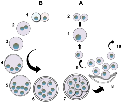
The entry of Pneumocystis into the mammalian lung likely occurs during the first year after birth. The agent of infection is suspected to be airborne spores. Recent studies suggest that the cyst/ascus (containing eight spores) may be the agent of infection . After inhalation, the spores ultimately take residence in the terminal portion of the respiratory tree, the alveoli. Neither the mechanism of migration to the alveoli nor the form in which the organism arrives in the alveoli (intact ascus or individual spores) is known. (A) Asexual phase: Haploid trophic forms are thought to replicate asexually by binary fission, whereby the nuclear content is duplicated (1) along with cellular contents that (2) divide into two haploid trophic forms. (B) Sexual phase: Two presumptive mating types conjugate (1), undergo karyogamy (2), and produce a diploid zygote (3) that progresses through meiosis to produce four haploid nuclei (4) followed by an additional mitosis to produce eight nuclei (5). The nuclei are packaged into spores by invagination of the ascus cell membranes (6) to produce eight double-membrane spores (7). After completion, excystment occurs via a protunicate release by unknown mechanisms (8). The released spores become the vegetative forms that can then undergo asexual (9) or sexual replication with a presumed opposite mating type (10). The mechanism of exit out of the lung and the life cycle form that transits into the environment are unknown (the life cycle was composed using SmartDraw 10, San Diego, California).
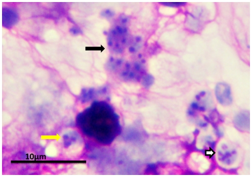
Clusters of P. murina from a homogenate of an infected mouse lung were dropped on glass slides and stained with a rapid Wright-Giemsa. The black arrow points to a cluster of trophic forms. The white arrow indicates a mature cyst. The yellow arrow indicates an immature cyst with only three nuclei present in this section. The magnification bar represents 10 um. The micrograph was taken with an Olympus BH2 microscope and DP-72 digital camera.
References
-
- Cushion MT. Pneumocystis pneumonia. In: Merz WG, Hay RJ, editors. Topley & Wilson's medical mycology. Washington (D.C.): Hodder Arnold. Distributed by ASM Press; 2005. pp. 763–806.
-
- Redhead SA, Cushion MT, Frenkel JK, Stringer JR. Pneumocystis and Trypanosoma cruzi: nomenclature and typifications. J Eukaryot Microbiol. 2006;53:2–11. - PubMed
-
- Hibbett DS, Binder M, Bischoff JF, Blackwell M, Cannon PF, et al. A higher-level phylogenetic classification of the Fungi Mycol Res. 2007;111:509–547. - PubMed
-
- Limper AH. Pneumocystis nomenclature. Clin Infect Dis. 2006;42:1210–1211. - PubMed
-
- Hawksworth DL. Responsibility in naming pathogens: the case of Pneumocystis jirovecii, the causal agent of pneumocystis pneumonia. Lancet Infect Dis. 2007;7:3–5. - PubMed
Publication types
MeSH terms
Grants and funding
LinkOut - more resources
Full Text Sources
Other Literature Sources
