. 2010 Apr;11(2):91-102.
doi: 10.2174/138920210790886835.

Genetic interference: don't stand so close to me

Affiliations

Genetic interference: don't stand so close to me

Luke E Berchowitz et al. Curr Genomics. 2010 Apr.

Abstract

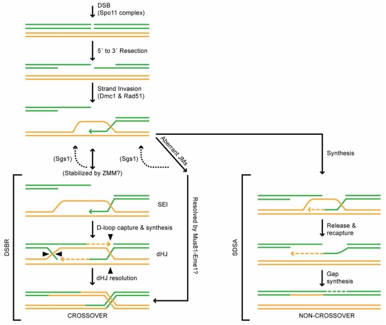
Meiosis is a dynamic process during which chromosomes undergo condensation, pairing, crossing-over and disjunction. Stringent regulation of the distribution and quantity of meiotic crossovers is critical for proper chromosome segregation in many organisms. In humans, aberrant crossover placement and the failure to faithfully segregate meiotic chromosomes often results in severe genetic disorders such as Down syndrome and Edwards syndrome. In most sexually reproducing organisms, crossovers are more evenly spaced than would be expected from a random distribution. This phenomenon, termed interference, was first reported in the early 20(th) century by Drosophila geneticists and has been subsequently observed in a vast range of organisms from yeasts to humans. Yet, many questions regarding the behavior and mechanism of interference remain poorly understood. In this review, we examine results new and old, from a wide range of organisms, to begin to understand the progress and remaining challenges to understanding the fundamental unanswered questions regarding genetic interference.

Keywords: Double-strand break; Meiosis; Pch2.; Rec8; chromosome Spo11; crossover; recombination; synaptonemal complex.


Figures

Fig. (1)
Fig. (1)
The DSBR and SDSA meiotic recombination models. Single strands of DNA are shown as either green (parent 1) or yellow (parent 2) rods. The Spo11 complex initiates programmed DSBs. DSBs are resected 5’ to 3’ to produce single ssDNA tails. ssDNA tails invade the homologous template which is aided by the ssDNA filament forming proteins Dmc1 and Rad51. At this stage, intermediates can undergo DSBR (left), which is thought to produce primarily COs or SDSA (right), which only produces NCOs. Also shown is a pathway (center) describing aberrant JMs, that are hypothesized to either be resolved back to the strand invasion stage by Sgs1 or resolved as COs by the Mus81-Eme1 heterodimer [52]. In the DSBR pathway, strand invasion complexes are stabilized (possibly by ZMM proteins) to form SEIs. Prior to stabilization, Sgs1 could wire SEIs back to the strand invasion stage [52, 89]. The displaced strand, called a D-loop is captured by the resected break of opposite homolog and subsequent DNA synthesis results in a dHJ intermediate. This intermediate is resolved as a CO upon appropriate resolution of the two HJs. in the SDSA pathway, the invading strand dissociates after a patch of DNA synthesis. This strand then re-anneals to the original parent, resulting in repair of the DSB and a patch of heteroduplex DNA. This pathway is always resolved as a NCO, but it can result in GC.
Fig. (2)
Fig. (2)
Interference Models. The left panel depicts the beam-film demonstration of the mechanical stress model proposed by Kleckner et al. [59]. The beam (chromosomal axis; green), film (chromatin fiber; grey), flaws (CO precursors; black dots). Diagrams depicting the stress level are shown under each beam in which the x axis represents beam position and stress level on the y. The center panel depicts the polymerization model proposed by King and Mortimer [75]. Chromatids are shown in green (parent 1) and yellow (parent 2). Small light blue circles represent recombination precursors and CO designates are shown as larger circles marked with ‘CO’. The interference polymer is shown as a large arrow emanating from CO sites, and CO precursors removed by the polymer are shown to the right accompanied with a dashed arrow. The right panel depicts the counting model proposed by Foss et al. [78]. Chromatids are shown in green (parent 1) and yellow (parent 2). Small light blue circles represent recombination precursors and CO designates are shown as larger circles marked with ‘CO’. In this diagram, m=3 and intervening NCOs between COs are outlined in a red box.

References

    1. Zickler D, Kleckner N. The leptotene-zygotene transition of meiosis. Annu. Rev. Genet. 1998;32:619–697. - PubMed
    1. Sturtevant AH. A Third Group of Linked Genes in Drosophila Ampelophila. Science. 1913;37:990–992. - PubMed
    1. Sturtevant AH. The behavior of chromosomes as studied through linkage. Z. Induct. Abstammungs-Vererbungsl. 1915;13:234–287.
    1. Borner GV, Kleckner N, Hunter N. Crossover/noncrossover differentiation, synaptonemal complex formation, and regulatory surveillance at the leptotene/zygotene transition of meiosis. Cell. 2004;117:29–45. - PubMed
    1. Jones GH. The control of chiasma distribution. Symp. Soc. Exp. Biol. 1984;38:293–320. - PubMed

LinkOut - more resources