Review
. 2010 Apr-Jun;6(2):76-87.
doi: 10.4161/org.6.2.10375.

The regulation of organ size in Drosophila: physiology, plasticity, patterning and physical force

Affiliations
Review

The regulation of organ size in Drosophila: physiology, plasticity, patterning and physical force

Alexander W Shingleton. Organogenesis. 2010 Apr-Jun.

Abstract

The correct regulation of organ size is a fundamental developmental process, the failure of which can compromise organ function and organismal integrity. Consequently, the mechanisms that regulate organ size have been subject to intense research. This research has highlighted four classes of mechanism that are involved in organ size regulation: physiology, plasticity, patterning and physical force. Nevertheless, how these mechanisms are integrated and converge on the cellular process that regulate organ growth is unknown. One group of animals where this integration is beginning to be achieved is in the insects. Here, I review the different mechanisms that regulate organ size in insects, and describe our current understanding of how these mechanisms interact. The genes and hormones involved are remarkably conserved in all animals, so these studies in insects provide a precedent for future research on organ size regulation in mammals.

Keywords: adhesion; hormone; hypoplasia; imaginal disc; insect; insulin; morphogens; size regulation.


Figures

Figure 1
Figure 1
The life cycle of Drosophila melanogaster. Larvae molt through three larval instars before metamorphosing into their adult form. Drosophila is a holometabolous insect and its adult organs develop as imaginal discs within the larva. Inset shows approximate positions of imaginal discs. Colors relate disc to corresponding adult organ.
Figure 2
Figure 2
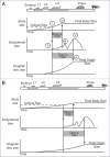
The physiological regulation of body and organ size in Drosophila. (A) Larvae grow until they reach a critical size at the beginning of the third larval instar (i), which initiates a hormonal cascade that ultimately causes the release of ecdysteroids (ii). When the ecdysteroids rise above a certain level (iii) it causes the larvae to stop feeding and begin wandering (iv) stopping body growth. Subsequent peaks in the ecdysteroid titre (v) cause pupation and the cessation of organ growth (vi). (B) A reduction in nutrition slows growth and delays attainment of critical size, extending total developmental time. Attainment of critical size initiates the same hormonal cascade that brings about the cessation of body and imaginal disc growth. The temporal dynamics of this cascade are unaffected by nutrition. Slow growth of the body and imaginal discs now reduces the amount of growth they can achieve during their TGPs, reducing final body and organ size. Hormones other than ecdsyteroids may be involved in the cessation of disc growth. L1–L3, first to third larval instar; TGP, terminal growth period. Adapted from ref. .
Figure 3
Figure 3
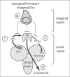
A model of critical size regulation in Drosophila. Critical size is regulated by larval and imaginal signals. The larvae signals comprise a (i) nutritional/size signal from the IPCs, and (ii) a temporal signal from the PTTH-producing neurons. The imaginal signal (iii) is inhibitory and may affect the action of dILPs or PTTH on the PG, or inhibit ecdysteroidgenesis directly. It is the balance of these signals that ultimately regulates the release of ecdysteroids (iv).
Figure 4
Figure 4
The II S and TOR-signaling pathway in Drosophila. The II S pathway is shown in cyan, the TOR-signaling pathway is shown in magenta, and the AMPK signaling pathway is shown in green. Variation in nutrition influences the release of dILPs, possibly by the action of sNPFs and AKH. dILPs bind to Inr which initiates a signal transduction cascade involving the phosphorylation of multiple intermediate proteins. Downstream growth affectors include dFOXO, which is deactivated by II S via phosphorylation by AKT, and S6K, which is activated by II S via PDK1. S6K is also a target of TOR, which also restricts the effects of dFOXO by inhibiting one of dFOXO’s transcriptional targets, 4EBP. TOR is also regulated indirectly by II S via the action of AKT on TSC1/2. TOR also responds to amino acids, by an unkown mehchanism, and glucose, via the AMPK pathway. Both FOXO and TOR regulate the activity of multiple growth inhibitors and promoters, respectively. Data from references 37, 43 and 50. Dotted lines are putative relationships.
Figure 5
Figure 5
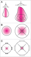
The autonomous regulation of organ size. (A) Day and Lawrence Model. The expression of a morphogen (e.g., Dpp) along the axis of an organ (e.g., the wing) establishes a morphogen gradient perpendicular to the axis (left). Growth stops when the morphogen gradient becomes sufficiently flat (right). (B) Shraiman model., A growth factor at the center of an organ promotes growth. Growth at the center stops when the positive effects of the growth factors are matched by the negative effects of compression (gray arrows), and at the periphery when the tissue grows beyond the range of the growth factor. (C) Aegerter-Wilmsen Model: A growth factor at the center of the organ promotes growth, which causes stretch at the periphery (black arrows), which in turn promotes growth at the periphery. Peripheral growth does not completely remove stretch, causing compression at the center of the organ. Growth stops when compression at the center overcomes the effects of the growth factor, eliminating additional stretch at the periphery (gray arrows) and stopping growth there also. Magenta area indicates morphogen. Adapted from ref. .
Figure 6
Figure 6
A model of the Fat/Hippo regulation of growth. Dpp signaling may affect signaling through the pathway via its effects on Dachsous and Fat. Wingless signaling may affect the activity of Yorkie through Vestigial’s interaction with Scalloped, a Yorkie co-factor. How Vestigial affects the interaction between Yorkie and Scalloped is, however, unclear. Mechanical forces may affect the activity of Hippo through their effect on Merlin and Expanded. Solid lines indicate protein-protein binding, solid arrows indicate biochemical interactions, dotted arrows indicate genetic interactions, gray arrow indicates hypothetical interaction. Data from references 96, 99 and 103.

Similar articles

Cited by

References

    1. Nijhout HF. Physiological control of molting in insects. Am Zool. 1981;21:631–640.
    1. Riddiford LM. Hormones and Drosophila development. In: Bate M, Martinez Arias A, editors. The Development of Drosophila melanogaster. Cold Spring Harbor: Cold Spring Harbor Press; 1993. pp. 899–939.
    1. Truman JW, Riddiford LM. Physiology of insect rhythms 3. The temporal organization of the endocrine events underlying pupation of the tobacco hornworm. J Exp Biol. 1974;60:371–382. - PubMed
    1. Kawasaki H. Transition from larva to pupa: morphogenesis, cell proliferation and protein synthesis in Bombyx wing disc. Inv Repro Devel. 1998;34:101–108.
    1. Ohtaki T, Yamanaka F, Sakurai S. Differential timing of pupal commitment in various tissues of the silkworm, Bombyx mori. J Insect Physiol. 1986;32:635.

Publication types

LinkOut - more resources