

Non-β-cell progenitors of β-cells in pregnant mice
- PMID: 20885859
- PMCID: PMC2901816
- DOI: 10.4161/org.6.2.10374
Non-β-cell progenitors of β-cells in pregnant mice
Abstract
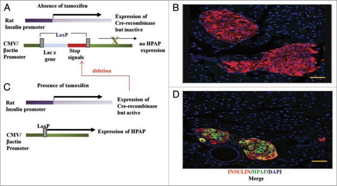
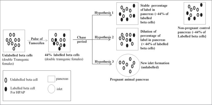
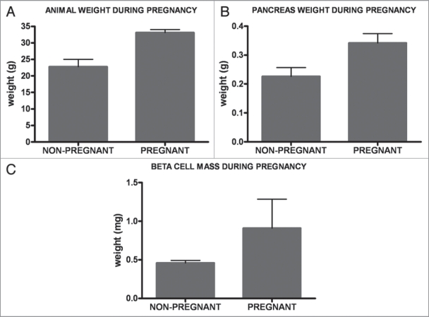
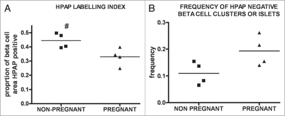
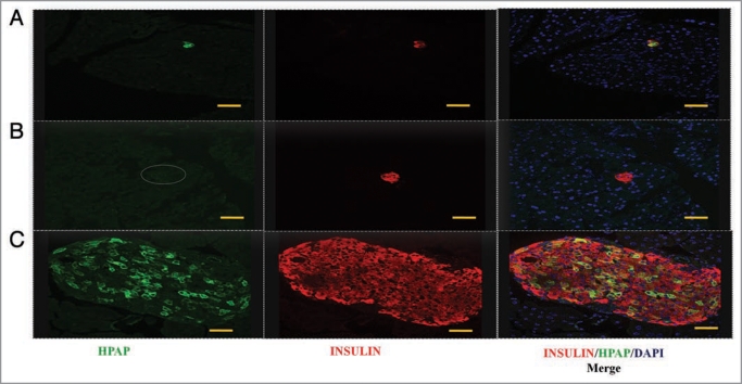
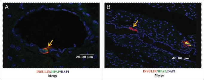
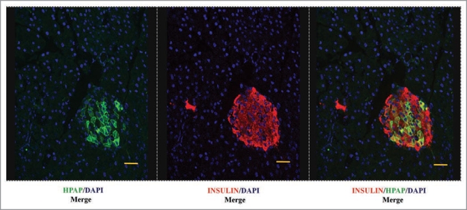
Pregnancy is a normal physiological condition in which the maternal β-cell mass increases rapidly about two-fold to adapt to new metabolic challenges. We have used a lineage tracing of β-cells to analyse the origin of new β-cells during this rapid expansion in pregnancy. Double transgenic mice bearing a tamoxifen-dependent Cre-recombinase construct under the control of a rat insulin promoter, together with a reporter Z/AP gene, were generated. Then, in response to a pulse of tamoxifen before pregnancy, β-cells in these animals were marked irreversibly and heritably with the human placental alkaline phosphatase (HP AP). First, we conclude that the lineage tracing system was highly specific for β-cells. Secondly, we scored the proportion of the β-cells marked with HP AP during a subsequent chase period in pregnant and non-pregnant females. We observed a dilution in this labeling index in pregnant animal pancreata, compared to nonpregnant controls, during a single pregnancy in the chase period. To extend these observations we also analysed the labeling index in pancreata of animals during the second of two pregnancies in the chase period. The combined data revealed statistically-significant dilution during pregnancy, indicating a contribution to new beta cells from a non-β-cell source. Thus for the first time in a normal physiological condition, we have demonstrated not only β-cell duplication, but also the activation of a non-β-cell progenitor population. Further, there was no transdifferentiation of β-cells to other cell types in a two and half month period following labeling, including the period of pregnancy.
Keywords: non-β-cell progenitor; pancreas; pregnancy; β-cell duplication; β-cells.
Figures
Similar articles
-
Transdifferentiation of pancreatic ductal cells to endocrine beta-cells.Biochem Soc Trans. 2008 Jun;36(Pt 3):353-6. doi: 10.1042/BST0360353. Biochem Soc Trans. 2008. PMID: 18481956
-
Genetic lineage tracing of beta cell neogenesis.Methods Mol Biol. 2012;933:317-22. doi: 10.1007/978-1-62703-068-7_21. Methods Mol Biol. 2012. PMID: 22893417
-
Preexisting pancreatic acinar cells contribute to acinar cell, but not islet beta cell, regeneration.J Clin Invest. 2007 Apr;117(4):971-7. doi: 10.1172/JCI29988. J Clin Invest. 2007. PMID: 17404620 Free PMC article.
-
Centroacinar cells: At the center of pancreas regeneration.Dev Biol. 2016 May 1;413(1):8-15. doi: 10.1016/j.ydbio.2016.02.027. Epub 2016 Mar 8. Dev Biol. 2016. PMID: 26963675 Free PMC article. Review.
-
Pancreatic acinar-to-beta cell transdifferentiation in vitro.Front Biosci. 2008 May 1;13:5824-37. doi: 10.2741/3119. Front Biosci. 2008. PMID: 18508625 Review.
Cited by
-
Development and regeneration in the endocrine pancreas.ISRN Endocrinol. 2012;2012:640956. doi: 10.5402/2012/640956. Epub 2012 Dec 27. ISRN Endocrinol. 2012. PMID: 23326678 Free PMC article.
-
Impaired proliferation of pancreatic beta cells, by reduced placental growth factor in pre-eclampsia, as a cause for gestational diabetes mellitus.Cell Prolif. 2015 Apr;48(2):166-74. doi: 10.1111/cpr.12164. Epub 2015 Jan 16. Cell Prolif. 2015. PMID: 25594238 Free PMC article.
-
Autonomous interconversion between adult pancreatic α-cells and β-cells after differential metabolic challenges.Mol Metab. 2016 May 10;5(7):437-448. doi: 10.1016/j.molmet.2016.05.001. eCollection 2016 Jul. Mol Metab. 2016. PMID: 27408770 Free PMC article.
-
Tamoxifen-Induced Cre-loxP Recombination Is Prolonged in Pancreatic Islets of Adult Mice.PLoS One. 2012;7(3):e33529. doi: 10.1371/journal.pone.0033529. Epub 2012 Mar 28. PLoS One. 2012. PMID: 22470452 Free PMC article.
-
Contribution of a non-β-cell source to β-cell mass during pregnancy.PLoS One. 2014 Jun 18;9(6):e100398. doi: 10.1371/journal.pone.0100398. eCollection 2014. PLoS One. 2014. PMID: 24940737 Free PMC article.
References
-
- Karnick SK, Chen H, McLean GW, Heit JJ, Gu X, Zhang AY, et al. Menin controls growth of pancreatic β-cells in pregnant mice and promotes gestational diabetes mellitus. Science. 2007;318:806–809. - PubMed
-
- Parsons JA, Bartke A, Sorenson RL. Number and size of islets of Langerhans in pregnant, human growth hormone-expressing transgenic, and pituitary dwarf mice: effect of lactogenic hormones. Endocrinology. 1995;136:2013–2021. - PubMed
-
- Scaglia L, Smith FE, Bonner-Weir S. Apoptosis contributes to the involution of β-cell mass in the post partum rat pancreas. Endocrinology. 1995;136:5461–5468. - PubMed
-
- Stanger BZ, Tanaka AJ, Melton DA. Organ size is limited by the number of embryonic progenitor cells in the pancreas but not the liver. Nature. 2007;445:886–891. - PubMed
-
- Parsons JA, Brelje TC, Sorenson RL. Adaptation of islets of Langerhans to pregnancy: increased islet cell proliferation and insulin secretion correlates with the onset of placental lactogen secretion. Endocrinology. 1992;130:1459–1466. - PubMed
Publication types
MeSH terms
Substances
Grants and funding
LinkOut - more resources
Full Text Sources
Medical
Molecular Biology Databases
Research Materials
Miscellaneous