

Case Reports
doi: 10.1155/2010/624825.
Epub 2010 Sep 15.
Laparoscopic retrieval of a peritoneal mouse
Affiliations
- PMID: 20885943
- PMCID: PMC2946616
- DOI: 10.1155/2010/624825
Item in Clipboard
Case Reports
Laparoscopic retrieval of a peritoneal mouse
Case Rep Med.
2010.
Abstract
A 67-year-old Caucasian male was referred by the urology service with a history of incomplete bowel emptying. He complained of tenesmus. MRI scan suggested a leiomyoma lying anterior to the rectum. He underwent examination under anaesthesia and attempted endorectal ultrasound and biopsy. However, the lesion seemed to migrate cranially and was impalpable. At laparoscopy, a mobile, unattached, 5.5 × 5 × 3.5, cream-coloured 'egg was retrieved from the retrovesical space. Histology confirmed a hyalinised fibrocollagenous lesion lined with mesothelium. A comprehensive review of the literature is presented.
Figures
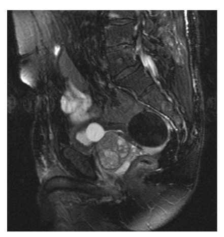
T2-weighted (fat-suppressed) sagittal image of the pelvis reveals a 5.5 cm retrovesical lesion lying anterior to the rectum. It is well circumscribed. There is no pelvic sidewall lymphadenopathy.
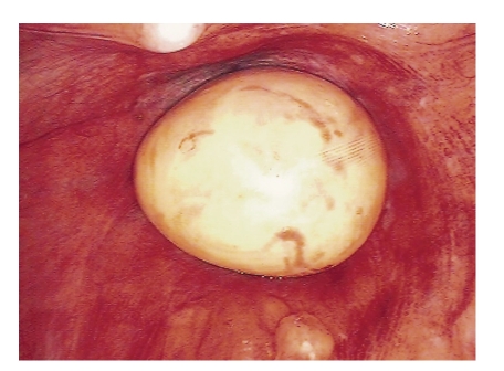
A cream-coloured ovoid mass is seen anterior to the rectum with the tip of the urethral catheter visible anterior to it.
References
-
- Koga K, Hiroi H, Osuga Y, Nagai M, Yano T, Taketani Y. Autoamputated adnexa presents as a peritoneal loose body. Fertility and Sterility. 2010;93(3):967–968. - PubMed
-
- Mohri T, Kato T, Suzuki H. A giant peritoneal loose body: report of a case. American Surgeon. 2007;73(9):895–896. - PubMed
-
- Ghosh P, Strong C, Naugler W, Haghighi P, Carethers JM. Peritoneal mice implicated in intestinal obstruction: report of a case and review of the literature. Journal of Clinical Gastroenterology. 2006;40(5):427–430. - PubMed
-
- Asabe K, Maekawa T, Yamashita Y, Shirakusa T. Endoscopic extraction of a peritoneal loose body: a case report of an infant. Pediatric Surgery International. 2005;21(5):388–389. - PubMed
-
- Ohgitani D, Kani H, Matsuki M, Kanazawa S, Narabayashi I. A case of giant peritoneal loose body: usefulness of wide window width CT. Nippon Acta Radiologica. 2004;64(4):223–224. - PubMed
Publication types
LinkOut - more resources
Full Text Sources
