

Sets of RNA repeated tags and hybridization-sensitive fluorescent probes for distinct images of RNA in a living cell
- PMID: 20885944
- PMCID: PMC2946342
- DOI: 10.1371/journal.pone.0013003
Sets of RNA repeated tags and hybridization-sensitive fluorescent probes for distinct images of RNA in a living cell
Abstract
Background: Imaging the behavior of RNA in a living cell is a powerful means for understanding RNA functions and acquiring spatiotemporal information in a single cell. For more distinct RNA imaging in a living cell, a more effective chemical method to fluorescently label RNA is now required. In addition, development of the technology labeling with different colors for different RNA would make it easier to analyze plural RNA strands expressing in a cell.
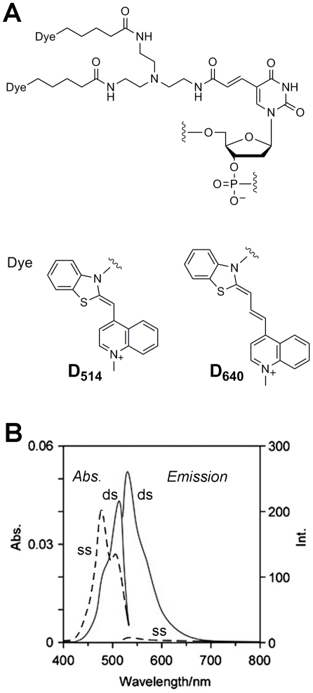
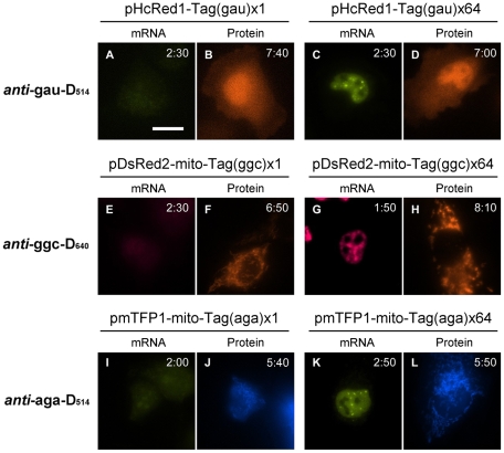
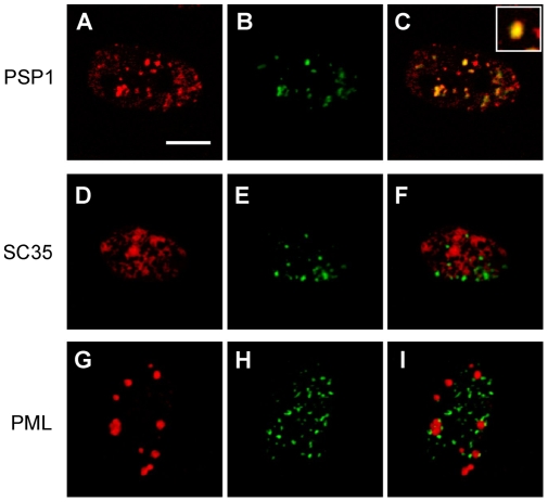
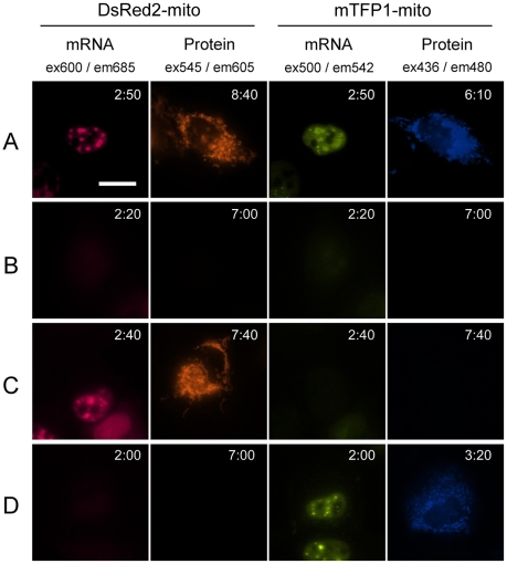
Methodology/principal findings: Tag technology for RNA imaging in a living cell has been developed based on the unique chemical functions of exciton-controlled hybridization-sensitive oligonucleotide (ECHO) probes. Repetitions of selected 18-nucleotide RNA tags were incorporated into the mRNA 3'-UTR. Pairs with complementary ECHO probes exhibited hybridization-sensitive fluorescence emission for the mRNA expressed in a living cell. The mRNA in a nucleus was detected clearly as fluorescent puncta, and the images of the expression of two mRNAs were obtained independently and simultaneously with two orthogonal tag-probe pairs.
Conclusions/significance: A compact and repeated label has been developed for RNA imaging in a living cell, based on the photochemistry of ECHO probes. The pairs of an 18-nt RNA tag and the complementary ECHO probes are highly thermostable, sequence-specifically emissive, and orthogonal to each other. The nucleotide length necessary for one tag sequence is much shorter compared with conventional tag technologies, resulting in easy preparation of the tag sequences with a larger number of repeats for more distinct RNA imaging.
Conflict of interest statement
Figures
Similar articles
-
Poly(A)+ Sensing of Hybridization-Sensitive Fluorescent Oligonucleotide Probe Characterized by Fluorescence Correlation Methods.Int J Mol Sci. 2021 Jun 16;22(12):6433. doi: 10.3390/ijms22126433. Int J Mol Sci. 2021. PMID: 34208525 Free PMC article.
-
Exciton-controlled fluorescence: application to hybridization-sensitive fluorescent DNA probe.Nucleic Acids Symp Ser (Oxf). 2009;(53):49-50. doi: 10.1093/nass/nrp025. Nucleic Acids Symp Ser (Oxf). 2009. PMID: 19749254
-
Hybridization-sensitive fluorescent oligonucleotide probe conjugated with a bulky module for compartment-specific mRNA monitoring in a living cell.Bioconjug Chem. 2015 Mar 18;26(3):412-7. doi: 10.1021/acs.bioconjchem.5b00090. Epub 2015 Mar 2. Bioconjug Chem. 2015. PMID: 25710491
-
ECHO probes: a concept of fluorescence control for practical nucleic acid sensing.Chem Soc Rev. 2011 Dec;40(12):5815-28. doi: 10.1039/c1cs15025a. Epub 2011 Jun 10. Chem Soc Rev. 2011. PMID: 21660343 Review.
-
Development of an effective protein-labeling system based on smart fluorogenic probes.J Biol Inorg Chem. 2019 Jun;24(4):443-455. doi: 10.1007/s00775-019-01669-y. Epub 2019 May 31. J Biol Inorg Chem. 2019. PMID: 31152238 Review.
Cited by
-
ECHO-liveFISH: in vivo RNA labeling reveals dynamic regulation of nuclear RNA foci in living tissues.Nucleic Acids Res. 2015 Oct 30;43(19):e126. doi: 10.1093/nar/gkv614. Epub 2015 Jun 22. Nucleic Acids Res. 2015. PMID: 26101260 Free PMC article.
-
The dynamic pathway of nuclear RNA in eukaryotes.Nucleus. 2013 May-Jun;4(3):195-205. doi: 10.4161/nucl.24434. Epub 2013 Apr 11. Nucleus. 2013. PMID: 23580182 Free PMC article. Review.
-
Broad Applications of Thiazole Orange in Fluorescent Sensing of Biomolecules and Ions.Molecules. 2021 May 10;26(9):2828. doi: 10.3390/molecules26092828. Molecules. 2021. PMID: 34068759 Free PMC article. Review.
-
Development of Highly Fluorogenic Styrene Probes for Visualizing RNA in Live Cells.ACS Chem Biol. 2023 Jul 21;18(7):1523-1533. doi: 10.1021/acschembio.3c00141. Epub 2023 May 18. ACS Chem Biol. 2023. PMID: 37200527 Free PMC article.
-
Fluorescent triplex-forming DNA oligonucleotides labeled with a thiazole orange dimer unit.Artif DNA PNA XNA. 2013 Jan-Mar;4(1):19-27. doi: 10.4161/adna.24102. Epub 2013 Jan 1. Artif DNA PNA XNA. 2013. PMID: 23445822 Free PMC article.
References
-
- Perlette J, Tan W. Real-time monitoring of intracellular mRNA hybridization inside single living cells. Anal Chem. 2001;73:5544–5550. - PubMed
Publication types
MeSH terms
Substances
LinkOut - more resources
Full Text Sources
Other Literature Sources
Research Materials
