

Defining hypo-methylated regions of stem cell-specific promoters in human iPS cells derived from extra-embryonic amnions and lung fibroblasts
- PMID: 20885964
- PMCID: PMC2946409
- DOI: 10.1371/journal.pone.0013017
Defining hypo-methylated regions of stem cell-specific promoters in human iPS cells derived from extra-embryonic amnions and lung fibroblasts
Abstract
Background: Human induced pluripotent stem (iPS) cells are currently used as powerful resources in regenerative medicine. During very early developmental stages, DNA methylation decreases to an overall low level at the blastocyst stage, from which embryonic stem cells are derived. Therefore, pluripotent stem cells, such as ES and iPS cells, are considered to have hypo-methylated status compared to differentiated cells. However, epigenetic mechanisms of "stemness" remain unknown in iPS cells derived from extra-embryonic and embryonic cells.
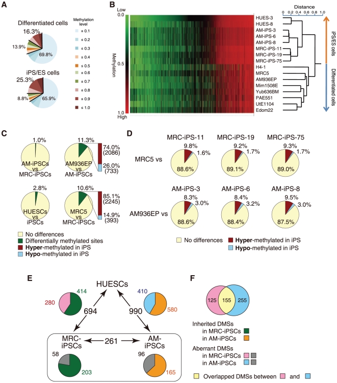
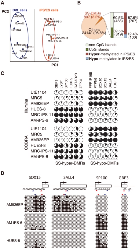
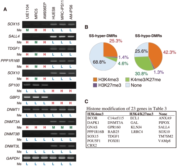
Methodology/principal findings: We examined genome-wide DNA methylation (24,949 CpG sites covering 1,3862 genes, mostly selected from promoter regions) with six human iPS cell lines derived from human amniotic cells and fetal lung fibroblasts as well as two human ES cell lines, and eight human differentiated cell lines using Illumina's Infinium HumanMethylation27. A considerable fraction (807 sites) exhibited a distinct difference in the methylation level between the iPS/ES cells and differentiated cells, with 87.6% hyper-methylation seen in iPS/ES cells. However, a limited fraction of CpG sites with hypo-methylation was found in promoters of genes encoding transcription factors. Thus, a group of genes becomes active through a decrease of methylation in their promoters. Twenty-three genes including SOX15, SALL4, TDGF1, PPP1R16B and SOX10 as well as POU5F1 were defined as genes with hypo-methylated SS-DMR (Stem cell-Specific Differentially Methylated Region) and highly expression in iPS/ES cells.
Conclusions/significance: We show that DNA methylation profile of human amniotic iPS cells as well as fibroblast iPS cells, and defined the SS-DMRs. Knowledge of epigenetic information across iPS cells derived from different cell types can be used as a signature for "stemness" and may allow us to screen for optimum iPS/ES cells and to validate and monitor iPS/ES cell derivatives for human therapeutic applications.
Conflict of interest statement
Figures
Similar articles
-
Investigating cellular identity and manipulating cell fate using induced pluripotent stem cells.Stem Cell Res Ther. 2012 Mar 8;3(2):8. doi: 10.1186/scrt99. Stem Cell Res Ther. 2012. PMID: 22405125 Free PMC article. Review.
-
Defining differentially methylated regions specific for the acquisition of pluripotency and maintenance in human pluripotent stem cells via microarray.PLoS One. 2014 Sep 24;9(9):e108350. doi: 10.1371/journal.pone.0108350. eCollection 2014. PLoS One. 2014. PMID: 25250679 Free PMC article.
-
Differential methylation of tissue- and cancer-specific CpG island shores distinguishes human induced pluripotent stem cells, embryonic stem cells and fibroblasts.Nat Genet. 2009 Dec;41(12):1350-3. doi: 10.1038/ng.471. Epub 2009 Nov 1. Nat Genet. 2009. PMID: 19881528 Free PMC article.
-
Induction of pluripotency in human endothelial cells resets epigenetic profile on genome scale.Cell Cycle. 2010 Mar 1;9(5):937-46. doi: 10.4161/cc.9.5.10869. Epub 2010 Mar 6. Cell Cycle. 2010. PMID: 20160486
-
Epigenetic modifications in the embryonic and induced pluripotent stem cells.Gene Expr Patterns. 2018 Sep;29:1-9. doi: 10.1016/j.gep.2018.04.001. Epub 2018 Apr 4. Gene Expr Patterns. 2018. PMID: 29625185 Review.
Cited by
-
DNA methylation dynamics in human induced pluripotent stem cells over time.PLoS Genet. 2011 May;7(5):e1002085. doi: 10.1371/journal.pgen.1002085. Epub 2011 May 26. PLoS Genet. 2011. PMID: 21637780 Free PMC article.
-
Role of Cripto-1 during epithelial-to-mesenchymal transition in development and cancer.Am J Pathol. 2012 Jun;180(6):2188-200. doi: 10.1016/j.ajpath.2012.02.031. Epub 2012 Apr 26. Am J Pathol. 2012. PMID: 22542493 Free PMC article. Review.
-
Investigating cellular identity and manipulating cell fate using induced pluripotent stem cells.Stem Cell Res Ther. 2012 Mar 8;3(2):8. doi: 10.1186/scrt99. Stem Cell Res Ther. 2012. PMID: 22405125 Free PMC article. Review.
-
Defining differentially methylated regions specific for the acquisition of pluripotency and maintenance in human pluripotent stem cells via microarray.PLoS One. 2014 Sep 24;9(9):e108350. doi: 10.1371/journal.pone.0108350. eCollection 2014. PLoS One. 2014. PMID: 25250679 Free PMC article.
-
A stable chimeric fibroblast growth factor (FGF) can successfully replace basic FGF in human pluripotent stem cell culture.PLoS One. 2015 Apr 7;10(4):e0118931. doi: 10.1371/journal.pone.0118931. eCollection 2015. PLoS One. 2015. PMID: 25850016 Free PMC article.
References
-
- Thomson JA, Itskovitz-Eldor J, Shapiro SS, Waknitz MA, Swiergiel JJ, et al. Embryonic stem cell lines derived from human blastocysts. Science. 1998;282:1145–1147. - PubMed
-
- Takahashi K, Tanabe K, Ohnuki M, Narita M, Ichisaka T, et al. Induction of pluripotent stem cells from adult human fibroblasts by defined factors. Cell. 2007;131:861–872. - PubMed
-
- Huangfu D, Osafune K, Maehr R, Guo W, Eijkelenboom A, et al. Induction of pluripotent stem cells from primary human fibroblasts with only Oct4 and Sox2. Nat Biotechnol. 2008;26:1269–1275. - PubMed
-
- Dimos JT, Rodolfa KT, Niakan KK, Weisenthal LM, Mitsumoto H, et al. Induced pluripotent stem cells generated from patients with ALS can be differentiated into motor neurons. Science. 2008;321:1218–1221. - PubMed
Publication types
MeSH terms
Substances
LinkOut - more resources
Full Text Sources
Other Literature Sources
