. 2010 Sep 24;5(9):e12969.
doi: 10.1371/journal.pone.0012969.

Curcumin protects intestinal mucosal barrier function of rat enteritis via activation of MKP-1 and attenuation of p38 and NF-κB activation

Affiliations

Curcumin protects intestinal mucosal barrier function of rat enteritis via activation of MKP-1 and attenuation of p38 and NF-κB activation

Wei-Bing Song et al. PLoS One. .

Abstract

Background: Intestinal mucosa barrier (IMB) dysfunction results in many notorious diseases for which there are currently few effective treatments. We studied curcumin's protective effect on IMB and examined its mechanism by using methotrexate (MTX) induced rat enteritis model and lipopolysaccharide (LPS) treated cell death model.

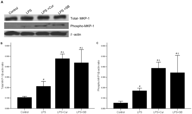
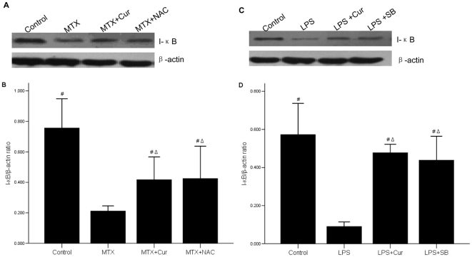
Methodology/principal findings: Curcumin was intragastrically administrated from the first day, models were made for 7 days. Cells were treated with curcumin for 30 min before exposure to LPS. Rat intestinal mucosa was collected for evaluation of pathological changes. We detected the activities of D-lactate and diamine oxidase (DAO) according to previous research and measured the levels of myeloperoxidase (MPO) and superoxide dismutase (SOD) by colorimetric method. Intercellular adhesion molecule-1 (ICAM-1), tumor necrosis factor α (TNF-α) and interleukin 1β (IL-1β) were determined by RT-PCR and IL-10 production was determined by ELISA. We found Curcumin decreased the levels of D-lactate, DAO, MPO, ICAM-1, IL-1β and TNF-α, but increased the levels of IL-10 and SOD in rat models. We further confirmed mitogen-activated protein kinase phosphatase-1 (MKP-1) was activated but phospho-p38 was inhibited by curcumin by western blot assay. Finally, NF-κB translocation was monitored by immunofluorescent staining. We showed that curcumin repressed I-κB and interfered with the translocation of NF-κB into nucleus.

Conclusions/significance: The effect of curcumin is mediated by the MKP-1-dependent inactivation of p38 and inhibition of NF-κB-mediated transcription. Curcumin, with anti-inflammatory and anti-oxidant activities may be used as an effective reagent for protecting intestinal mucosa barrier and other related intestinal diseases.


Conflict of interest statement

Competing Interests: The authors have declared that no competing interests exist.

Figures

Figure 1
Figure 1. Construction of MTX induced enteritis animal model.
Schedule of rat experimental procedure is shown by a diagram. Enteritis was induced in rats through peritoneal injection of MTX (20 mg/kg). Control group was only treated with saline. From the first day that the rat models were made, the drugs, curcumin or NAC, were intragastrically administrated with the specified dosage once a day for 7 days, then all rats were anesthetized and killed.
Figure 2
Figure 2. Effects of curcumin on DAO and D-lactate activities in small intestinal mucosa.
(A) MTX distinctly increased the level of D-lactate, but curcumin depressed the level of it. (B) The level of DAO were highter in MTX group compared to control group but lower in MTX+curcumin group The same results were found in MTX+NAC group. The levels of DAO and D-lactate of the small intestinal were detected by UV-spectrophotometry. *p = 0.000 vs. control group, #p = 0.000 vs. MTX group.
Figure 3
Figure 3. Effects of curcumin on MPO and ICAM-1 activity in vivo and in vitro.
(A) The level of MPO in small intestinal mucosa was increased after treated with curcumin or NAC. The level of MPO was determined using the colorimetric method. *p = 0.000 vs. control group, #p = 0.000 vs. MTX group. (B) ICAM-1 mRNA was down-regulated by curcumin or NAC in the intestinal mucosa of experiment rats. (C). ICAM-1 mRNA was down-regulated by curcumin or SB in LPS treated IEC-6 cells. The levels of ICAM-1 mRNA were detected by RT-PCR.
Figure 4
Figure 4. Effects of curcumin on the mRNA levels of TNF-α and IL-1β.
(A) The expressions of TNF-α and IL-1β mRNA in MTX group were increased, but it was inhibited after treating with curcumin or NAC. (B) The expressions of TNF-α and IL-1β in LPS treated IEC-6 cells were also increased, but it was depressed by curcumin or SB. The levels of TNF-α and IL-1β mRNA were detected by RT-PCR.
Figure 5
Figure 5. Effects of curcumin on the level of IL-10.
(A) The level of IL-10 was decreased in MTX group, but increased after treating with curcumin or NAC. *p = 0.000 vs. control group, #p = 0.000 vs. MTX group. (B) The level of IL-10 was decreased in the supernatant of LPS treated IEC-6 cells. Both Curcumin and SB promoted the expression of IL-10. *p = 0.000 vs. control group, #p = 0.000 vs. LPS group. The level of IL-10 was detected by ELISA.
Figure 6
Figure 6. Effects of curcumin on small intestinal SOD activity.
The level of SOD was inhibited by MTX, while elevated by treating with curcumin or NAC. The levels of SOD in the intestinal mucosa were determined using the colorimetric method. *p = 0.000 vs. control group, #p = 0.000 vs. MTX group.
Figure 7
Figure 7. Effect of curcumin on phosphorylation sate of p38.
(A) The expressions of total p38, phosphorylation sates of p38, Erk1/2, and JNK1/2 were all enhanced in MTX group. Only the phosphorylation sate of p38 was attenuated by treating with curcumin or NAC. The expressions of phosphorylated or total proteins were detected by western blot. (B) The bar graph was a clear reflection of the level of phosphorylation sate of p38 among four groups. *p = 0.000 vs. control group, #p = 0.000 vs. MTX group.
Figure 8
Figure 8. Effect of curcumin on both total MKP-1 and its phosphorylation state in LPS-stimulated IEC-6 cells.
(A) The expressions of total MKP-1 and phospho MKP-1 were highest in LPS+curcumin group and LPS+SB group, middle in LPS treated IEC-6 cells, and lowest in control group. The levels of total or phosphorylated proteins were detected by western blot. (B) (C) The bar graphs were a clear reflection of the level of total MKP-1 and phosphor-MKP-1 among four groups. #p<0.05 vs. control group; Δp = 0.00 vs. LPS group.
Figure 9
Figure 9. Effect of Curcumin on inhibition of I-κB degradation.
(A) The level of I-κB protein in the small intestinal tissue was decreased in MTX group, but increased by curcumin or NAC. (B) The bar graph was a clear reflection of the level of I-κB among four groups. #p = 0.000 vs. MTX group, Δp<0.01 vs. control group (C) The expression of I-κB protein was decreased in LPS group, but increased after treating curcumin or SB. (D) The expression of I-κB protein in LEC-6 cells was showed in bar graph. #p = 0.000 vs. LPS group, Δp<0.01 vs. control group. The level of I-κB protein was detected by western blot.
Figure 10
Figure 10. Effect of curcumin on blocking NF-κB translocation into nucleus.
(A) NF-κB p65 was located in the cytoplasm in control group (white arrow). (B) NF-κB p65 translocated to nucleus after stimulated by LPS (white arrow). (C) Curcumin attenuated the translocation of NF-κB p65 to nucleus (white arrow). (D) SB inhibited the translocation of NF-κB p65 into the nucleus (white arrow). These were observed by immunofluorescence. Magnification×400.

Similar articles

Cited by

References

    1. Aranow JS, Fink MP. Determinants of intestinal barrier failure in critical illness. Br J Anaesth. 1996;77:71–81. - PubMed
    1. Kalff JC, Hierholzer C, Tsukada K, Billiar TR, Bauer AJ. Hemorrhagic shock results in intestinal muscularis intercellular adhesion molecule (ICAM-1) expression, neutrophil infiltration,and smooth muscle dysfunction. Arch Orthop Trauma Surg. 1999;119:89–93. - PubMed
    1. Tibble JA, Sigthorsson G, Bridger S, Fagerhol MK, Bjarnason I. Surrogate markers of intestinal inflammation are predictive of relapse in patients with inflammatory bowel disease. Gastroenterology. 2000;119:15–22. - PubMed
    1. Ruh J, Vogel F, Schmidt E, Werner M, Klar E, et al. Effects of hydrogen peroxide scavenger Catalase on villous microcirculation in the rat small intestine in a model of inflammation bowel disease. Microvasc Res. 2000;59:329–337. - PubMed
    1. Nieto N, Torres MI, Fernández MI, Girón MD, Ríos A, et al. Experimental ulcerative colitis impairs antioxidant defense system in rat intestine. Dig Dis Sci. 2000;45:1820–1827. - PubMed

Publication types

MeSH terms