

Dermoscopy Clues in Pigmented Bowen's Disease
- PMID: 20886019
- PMCID: PMC2945663
- DOI: 10.1155/2010/464821
Dermoscopy Clues in Pigmented Bowen's Disease
Abstract
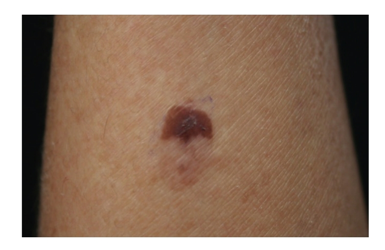
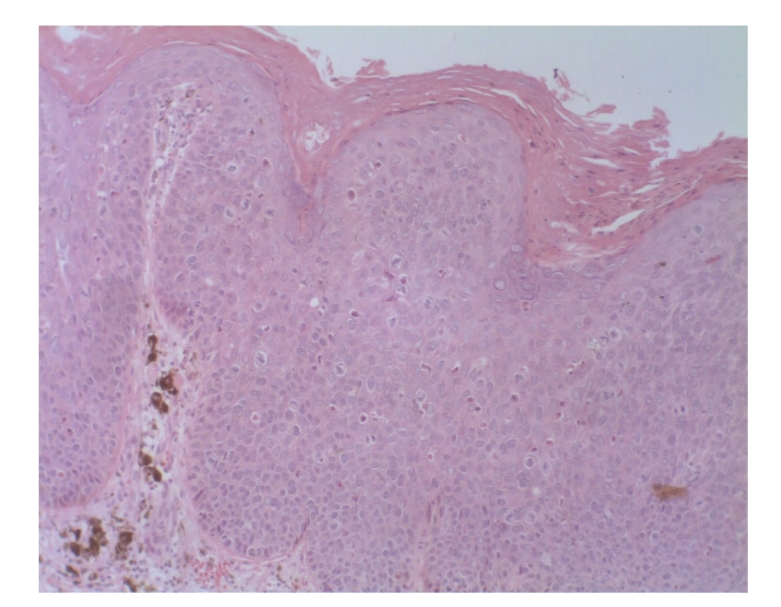
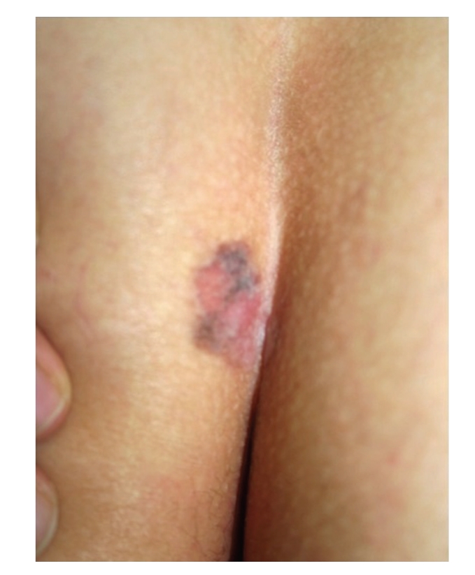
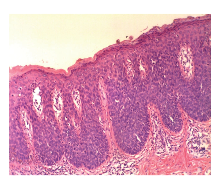
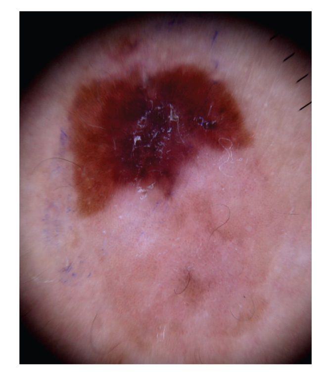
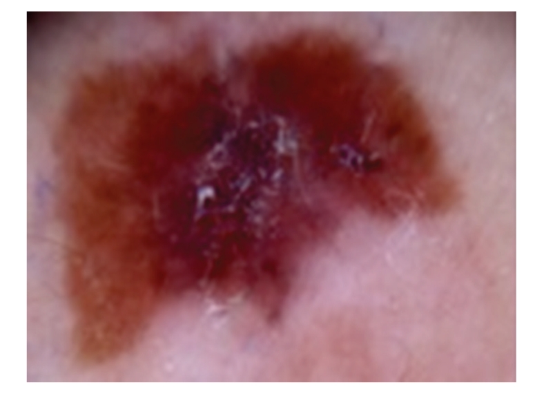
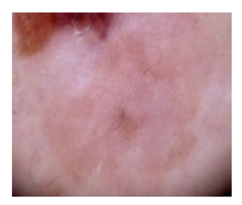
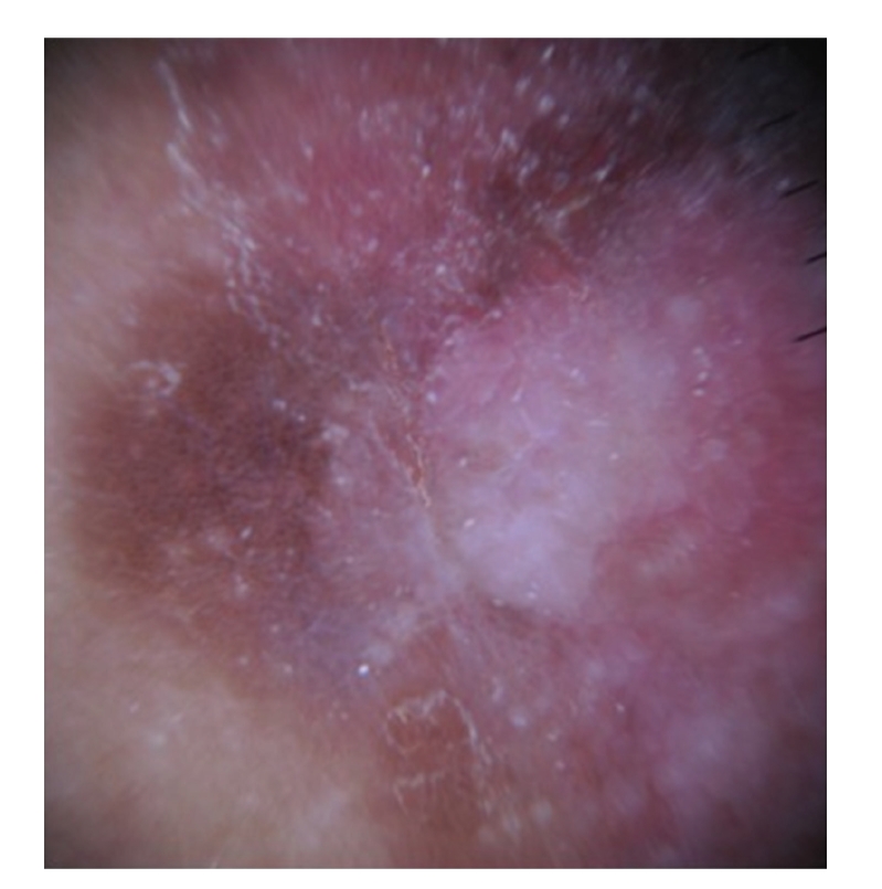
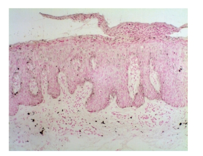
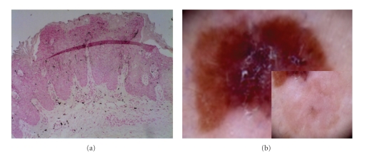
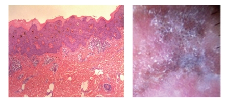
Pigmented tumors have similar clinical features that overlap and hamper diagnosis. Dermoscopy increases the diagnostic accuracy of doubtful melanocytic lesions and has been used as a noninvasive tool in the detection of pigmented lesions (PLs) like melanoma, basal cell carcinoma, and pigmented Bowen's disease (pBD). Our objective was to show the dermoscopic features of 2 cases of pBD and compare with the findings reported in the literature. Two dermoscopic images of biopsy proven pBD were retrospectively analyzed for dermoscopic patterns. Both cases showed brown regular globules, structureless brown and blue pigmentation, glomerular vessels, hypopigmented regression-like areas, and keratosis. These findings were similar to the cases reported previously. The dermoscopic diagnosis of pBD is based on the absence of criteria for a melanocytic lesion in the presence of glomerular vessels, regular brown globules and keratosis. Although pBD is rare, it should be included in the differential diagnosis of PLs, especially melanoma.
Figures


Similar articles
-
Pigmented Bowen's disease presenting with a "starburst" pattern.Dermatol Pract Concept. 2016 Oct 31;6(4):47-49. doi: 10.5826/dpc.0604a11. eCollection 2016 Oct. Dermatol Pract Concept. 2016. PMID: 27867748 Free PMC article.
-
Dermoscopy of Bowen's disease.Br J Dermatol. 2004 Jun;150(6):1112-6. doi: 10.1111/j.1365-2133.2004.05924.x. Br J Dermatol. 2004. PMID: 15214896
-
What's new in dermoscopy of Bowen's disease: two new dermoscopic signs and its differential diagnosis.Int J Dermatol. 2017 Oct;56(10):1022-1025. doi: 10.1111/ijd.13734. Epub 2017 Aug 22. Int J Dermatol. 2017. PMID: 28832993
-
Pigmented nonmelanoma skin cancers of the genital area: a diagnostic and therapeutic challenge - monocentric experience and review of the literature.Int J Dermatol. 2024 Nov;63(11):1477-1483. doi: 10.1111/ijd.17286. Epub 2024 Jun 5. Int J Dermatol. 2024. PMID: 38840323 Review.
-
Role of In Vivo Reflectance Confocal Microscopy in the Analysis of Melanocytic Lesions.Acta Dermatovenerol Croat. 2018 Apr;26(1):64-67. Acta Dermatovenerol Croat. 2018. PMID: 29782304 Review.
Cited by
-
A case of pigmented Bowen's disease.An Bras Dermatol. 2017 Jan-Feb;92(1):124-125. doi: 10.1590/abd1806-4841.20175381. An Bras Dermatol. 2017. PMID: 28225972 Free PMC article.
-
Race-Specific and Skin of Color Dermatoscopic Characteristics of Skin Cancer: A Literature Review.Dermatol Pract Concept. 2023 Oct 1;13(4 S1):e2023311S. doi: 10.5826/dpc.1304S1a311S. Dermatol Pract Concept. 2023. PMID: 37874992 Free PMC article. Review.
-
Dermoscopic Features of Pigmented Bowen Disease: A Multicenter Study on Behalf of the Ibero-Latin American College of Dermatology (CILAD).Dermatol Pract Concept. 2024 Apr 1;14(2):e2024086. doi: 10.5826/dpc.1402a86. Dermatol Pract Concept. 2024. PMID: 38810038 Free PMC article.
-
Multiple Pigmented Bowen's Disease: A Diagnostic and Therapeutic Dilemma.Case Rep Oncol Med. 2012;2012:342030. doi: 10.1155/2012/342030. Epub 2012 Oct 16. Case Rep Oncol Med. 2012. PMID: 23119201 Free PMC article.
-
Dermoscopic features of neoplasms in skin of color: A review.Int J Womens Dermatol. 2021 Jan 19;7(2):145-151. doi: 10.1016/j.ijwd.2020.11.009. eCollection 2021 Mar. Int J Womens Dermatol. 2021. PMID: 33937480 Free PMC article. Review.
References
-
- Zalaudek I, Citarella L, Soyer HP, Hofmann-Wellenhof R, Argenziano G. Dermoscopy features of pigmented squamous cell carcinoma: a case report. Dermatologic Surgery. 2004;30(4):539–540. - PubMed
-
- Zalaudek I, Argenziano G, Leinweber B, et al. Dermoscopy of Bowen’s disease. British Journal of Dermatology. 2004;150(6):1112–1116. - PubMed
-
- Krishnan R, Lewis A, Orengo IF, Rosen T. Pigmented bowen’s disease (squamous cell carcinoma in situ): a mimic of malignant melanoma. Dermatologic Surgery. 2001;27(7):673–674. - PubMed
-
- Stante M, De Giorgi V, Massi D, Chiarugi A, Carli P. Pigmented Bowen’s disease mimicking cutaneous melanoma: clinical and dermoscopic aspects. Dermatologic Surgery. 2004;30(4):541–544. - PubMed
-
- Soyer H, Argenziano G, Chimenti S, et al. Dermoscopy of Pigmented Skin Lesions. An Atlas Based on the Concensus Net Meeting on Dermoscopy. Milan, Italy: Edra; 2000.
Publication types
LinkOut - more resources
Full Text Sources
