. 2010 Sep 24;5(9):e13006.
doi: 10.1371/journal.pone.0013006.

Suppression of uPAR retards radiation-induced invasion and migration mediated by integrin β1/FAK signaling in medulloblastoma

Affiliations

Suppression of uPAR retards radiation-induced invasion and migration mediated by integrin β1/FAK signaling in medulloblastoma

Arun Kumar Nalla et al. PLoS One. .

Retraction in

Abstract

Background: Despite effective radiotherapy for the initial stages of cancer, several studies have reported the recurrence of various cancers, including medulloblastoma. Here, we attempt to capitalize on the radiation-induced aggressive behavior of medulloblastoma cells by comparing the extracellular protease activity and the expression pattern of molecules, known to be involved in cell adhesion, migration and invasion, between non-irradiated and irradiated cells.

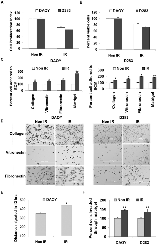
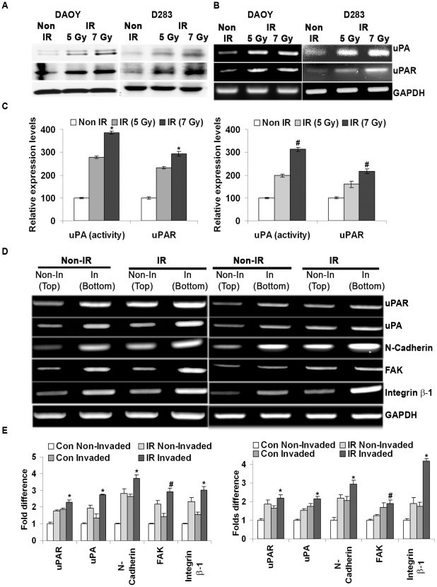
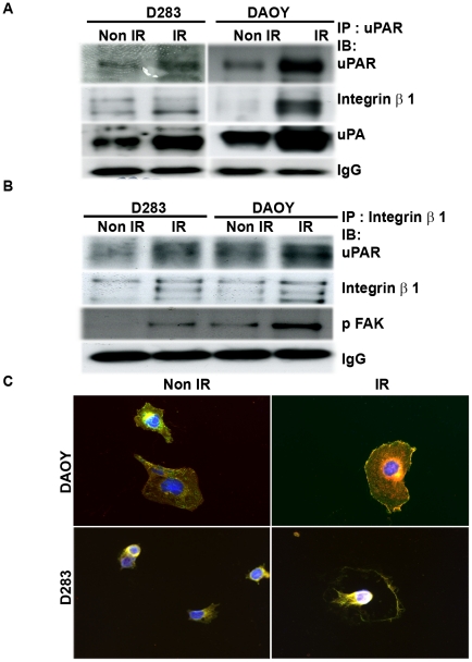
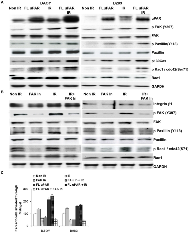
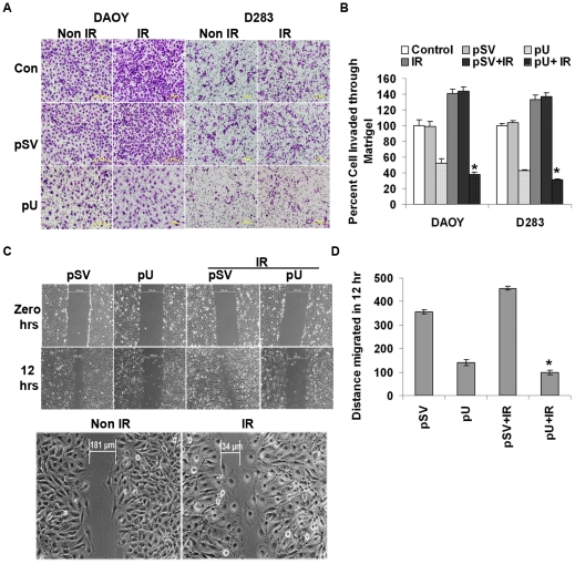
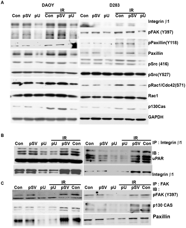
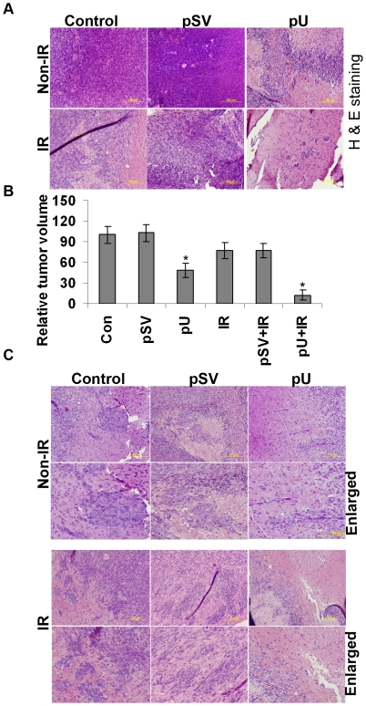
Methodology/principal findings: We identified an increase in invasion and migration of irradiated compared to non-irradiated medulloblastoma cells. RT-PCR analysis confirmed increased expression of uPA, uPAR, focal adhesion kinase (FAK), N-Cadherin and integrin subunits (e.g., α3, α5 and β1) in irradiated cells. Furthermore, we noticed a ∼2-fold increase in tyrosine phosphorylation of FAK in irradiated cells. Immunoprecipitation studies confirmed increased interaction of integrin β1 and FAK in irradiated cells. In addition, our results show that overexpression of uPAR in cancer cells can mimic radiation-induced activation of FAK signaling. Moreover, by inhibiting FAK phosphorylation, we were able to reduce the radiation-induced invasiveness of the cancer cells. In this vein, we studied the effect of siRNA-mediated knockdown of uPAR on cell migration and adhesion in irradiated and non-irradiated medulloblastoma cells. Downregulation of uPAR reduced the radiation-induced adhesion, migration and invasion of the irradiated cells, primarily by inhibiting phosphorylation of FAK, Paxillin and Rac-1/Cdc42. As observed from the immunoprecipitation studies, uPAR knockdown reduced interaction among the focal adhesion molecules, such as FAK, Paxillin and p130Cas, which are known to play key roles in cancer metastasis. Pretreatment with uPAR shRNA expressing construct reduced uPAR and phospho FAK expression levels in pre-established medulloblastoma in nude mice.

Conclusion/significance: Taken together, our results show that radiation enhances uPAR-mediated FAK signaling and by targeting uPAR we can inhibit radiation-activated cell adhesion and migration both in vitro and in vivo.


Conflict of interest statement

Competing Interests: The authors have declared that no competing interests exist.

Figures

Figure 1
Figure 1. Radiation reduced the survival rate but enhanced cell adhesion, migration and invasion of medulloblastoma cells.
Cells were irradiated with 7 Gy and the effect of radiation on cell proliferation rate and viability was determined by (A) MTT assay and (B) trypan blue exclusion assay. Mean values (± S.D, p<0.001) from three independent experiments were represented graphically. (C) Radiation enhanced the adhesion of cells to ECM components. 24 hrs after irradiating the cells with 7 Gy, the cells were detached from the plates and an equal number of cells (irradiated [IR] and non-irradiated [non-IR]) were plated on a 96-well culture plate coated either with type-I collagen (5 µg/mL), fibronectin (2 µg/mL), vitronectin (2 µg/mL) and matrigel (50 µg/mL). After 1 hr (DAOY) or 3 hrs (D283), the cells that attached to collagen, fibronectin and vitronectin were stained with Hema-3 and visualized under a light microscope. The number of cells attached to matrigel was quantified by MTT assay. The number of cells attached to various ECM components was counted and graphically represented. Bars represent the mean ± S.D values from three different experiments (* and ** represents p<0.05 and p<0.005, respectively, compared to the respective non-irradiated cells in each group). (D) Represents the spreading of cells induced upon binding of IR and non-IR DAOY and D283 cells to ECM components. (E) Wound healing migration assay was carried out on DAOY cells. Distance migrated by non-IR and IR cells was compared and represented in the graph (mean ± S.D value from three independent experiments; * represents p<0.05 compared to non-irradiated DAOY cells). (F) Invasiveness of DAOY and D283 cells (with and without radiation) was quantified by counting the number of cells that invaded through matrigel. Results of five different experiments are represented (± S.D. value, ** p<0.05 compared to respective non-irradiated cells).
Figure 2
Figure 2. Radiation enhanced extracellular protease activity and aggressiveness of the cancer cells.
(A) Medulloblastoma cells were irradiated with 7 Gy and serum starved for 24 hrs. The conditioned medium was collected and analyzed by SDS-PAGE gels either containing fibrinogen/plasminogen to determine the enzymatic activity of uPA. uPAR levels were detected in total cell lysates by immunoblotting with uPAR-specific antibodies. GAPDH antibody was used to confirm equal loading of the proteins in each lane. (B) Total RNA was isolated from the non-irradiated and irradiated cells, and transcript levels of uPA and uPAR were determined by RT-PCR analysis. (C) The enzymatic activity as determined by clear zone on the zymography gels were quantified by densitometry. Intensity of uPAR signals were quantified by densitometery and normalized with GAPDH. Graphical representation of values obtained from four independent experiments was presented with a mean ± S.D value, (*p<0.01 and # p<0.05 with reference to respective controls) (D) Translation modification of matrigel-invading cells (In) was compared between non-irradiated (non-IR) and irradiated (IR) cells. Total RNA was isolated from non-invading and matrigel-invading cells as described in Materials and Methods. RT-PCR analysis was carried out for transcript levels of integrin subunits, uPAR, uPA, N-Cadherin and FAK were determined using specific primers. GAPDH was used as a control. (E) The amplicon intensity was quantified and represented. Results from two different experiments are represented (± S.D. value, * p<0.05 and # p<0.01 with reference to non-invading controls).
Figure 3
Figure 3. Radiation induced uPAR/integrin β1/FAK interaction led to activation of FAK/Src/Rac signaling.
Co-immunoprecipitation assay was carried out to determine the interaction of uPAR with integrin ββ1. Cells were irradiated and detached from the culture plate using a cell stripper solution. Total cell lysates were immunoprecipitated with either uPAR or integrin β1. (A) Immunoprecipitates of uPAR were analyzed by immunoblotting with integrin β1 and uPA antibodies. (B) Immunoprecipitates of uPAR/integrin β1 were analyzed by western blotting with uPAR, FAK and pFAK (Y397). (C) Immunofluorescence assay was used to detect the localization of uPAR/integrin β1 in irradiated and non-irradiated cells. Cells seeded on 2-well chamber slides were irradiated with 7 Gy and incubated for 24 hrs. The cells were incubated with uPAR and integrin β1 antibodies followed by incubation with species-specific Alexa Fluor secondary antibodies. Immunofluorescence assay demonstrates the co-localization (yellow color) of uPAR and integrin β1 at the migratory front of irradiated cells.
Figure 4
Figure 4. uPAR overexpression enhances FAK signaling and cell invasion in medulloblastoma cells.
(A) DAOY and D283 cells were transiently transfected with full length uPAR (FL-uPAR) for 48 hrs. Transfected cells were either irradiated with 7 Gy or non-irradiated and incubated for another 24 hrs. Total cell lysates were immunoblotted with the indicated antibodies. (B) Inhibiting FAK phosphorylation reduced the radiation-induced activation of FAK signaling cascade and cell invasion. DAOY and D283 cells were treated with or without FAK inhibitor for 2 hrs and then subjected to radiation and incubated for another 24 hrs. Phosphorylation levels of FAK (Y397) and downstream signal molecules, such as Paxillin and Rac-1/Cdc42, were confirmed by immunoblotting the total cell lysates with specific antibodies. (C) FL-uPAR-transfected and/or FAK inhibited cells (either irradiated or non-irradiated) were subjected to matrigel invasion assay to determine the invasive potential of uPAR overexpression in cancer cells. The number of cells invaded per each treatment was counted and graphically presented. Results from three different experiments were complied and represented with a ± S.D mean value, p<0.05.
Figure 5
Figure 5. siRNA-mediated downregulation of uPAR reduced radiation-induced cell adhesion, invasion and migration.
A plasmid expressing siRNA against the uPAR gene (pU) was transiently transfected into DAOY and D283 cells. The efficiency of uPAR downregulation was confirmed by RT-PCR analysis using specific primers. (A1) After 36 hrs of transfection, the cells were either irradiated with 7 Gy or non-irradiated and incubated for another 24 hrs before total RNA was isolated. RT-PCR analysis was carried out to determine the effect of uPAR knockdown on the transcriptional levels of uPAR, integrin β1 and FAK. GAPDH was used as a loading control. (A2) Western analysis of the total cell lysates extracted from uPAR knockdown cells (with and with radiation) with uPAR and GAPDH antibodies. (B) Amplicon intensity was measured using densitometry. Data shown are the mean of three different experiments and the bars are ± S.D. (* p<0.05 with IR and # p<0.01 with control). (C) The effect of uPAR downregulation and radiation on the cells adhesive properties was determined by plating cells onto ECM component-coated plates. 1×10⁴ cells were allowed to adhere to plates coated (a) type-I collagen, (b) fibronectin or (c) vitronectin. After allowing the cells to attach for 1 hr (DAOY) or 3 hrs (D283), the wells were washed and stained with Hema-3. The number of adhered cells was counted under a light microscope and quantified data from three different experiments are represented graphically (mean ± S.D., * represents p value of <0.01).
Figure 6
Figure 6. Knockdown of uPAR reduced radiation-induced cell invasion and migration.
Knockdown of uPAR inhibited radiation-enhanced cell adhesion, invasion and migration by blocking uPAR/integrin β1/FAK interactions. After 48 hrs of pU transfection, the cells were either irradiated or non-irradiated before analysis for cell adhesion, invasion and migration potential. Cells were detached from the culture plate using cell stripper solution for further analysis. (A) 2×10⁵ cells were plated on matrigel-coated transwell inserts and incubated for 24 hrs to determine the invasive potential of irradiated and non-irradiated pU-transfected cancer cells. The matrigel-invading cells were stained with Hema-3 and visualized under a light microscope. (B) The number of invading cells was counted from five different fields and quantified; a comparison between various treatments is represented graphically. Percent cell invasion was measured from the mean obtained from three independent experiments and values shown are the mean ± S.D (* p<0.005 with reference to irradiated control). (C) Wound healing migration assay was carried out to determine the migratory ability of irradiated and non-irradiated pU-transfected cells. Transfected cells were grown until a monolayer formed and then a scratch was made using a 200-µL pipette tip. After thorough washing with PBS, the cells were either left non-irradiated or irradiated with 7 Gy. The distance migrated by the cells was monitored over a period of time by observation under a light microscope. (D) This was further quantified and data are represented graphically as a mean of three different experiments with ± S.D (* represents p<0.05).
Figure 7
Figure 7. uPAR knockdown reduces radiation-induced FAK signaling cascade by disrupting the interaction of focal adhesion molecules.
(A) Total cell lysates extracted from uPAR knockdown cells (either irradiated or non-irradiated) were analyzed by immunoblotting with integrin β1, phospho FAK (Y397), Paxillin, Rac-1/Cdc42 and p130Cas. GAPDH was used as a loading control. Radiation enhanced tyrosine phosphorylation of FAK was reduced by inhibiting the interaction of uPAR and integrin β1 in pU-transfected cells. Total cell lysates were subjected to immunoprecipitation with either integrin β1 or FAK. (B) The immunoprecipitates of integrin β1 were analyzed by western blotting with antibodies specific for uPAR and FAK. (C) The co-immunoprecipitates of FAK were analyzed by western blotting with antibodies specific for phospho FAK, Paxillin and p130CAS.
Figure 8
Figure 8. Effect of uPAR siRNA expressing plasmid treatment prior to radiation on pre-established intracranial tumor.
(A). Hematoxylin and eosin (H&E) staining performed on the paraffin embedded tissue sections of pre-established tumors from untreated and mice treated with either pU or pSV followed by with or without radiation. Experiments were carried out on five animals in each group. (B) Tumor volumes were quantified and represented graphically. (n = 5 with mean ± S.D; *p<0.05 with reference to non-irradiated control mice. (C) H&E staining of the brain section was carried out and representative pictures of brain sections showing the migratory fronts and metastatic bodies from control, pSV and pU (with and without radiation) treated mice are shown.

Similar articles

Cited by

References

    1. Crawford JR, MacDonald TJ, Packer RJ. Medulloblastoma in childhood: new biological advances. Lancet Neurol. 2007;6:1073–1085. - PubMed
    1. Howes TL, Buatti JM, Kirby PA, Carlisle TL, Ryken TC. Radiation induced adult medulloblastoma: a case report. J Neurooncol. 2006;80:191–194. - PubMed
    1. Chan MY, Teo WY, Seow WT, Tan AM. Epidemiology, management and treatment outcome of medulloblastoma in singapore. Ann Acad Med Singapore. 2007;36:314–318. - PubMed
    1. Feltbower RG, Picton S, Bridges LR, Crooks DA, Glaser AW, et al. Epidemiology of central nervous system tumors in children and young adults (0–29 years), Yorkshire, United Kingdom. Pediatr Hematol Oncol. 2004;21:647–660. - PubMed
    1. Ranger A, McDonald W, Bauman GS, Del MR. Effects of surgical excision and radiation on medulloblastoma cell invasiveness. Can J Neurol Sci. 2009;36:631–637. - PubMed

Publication types

MeSH terms

Substances