. 2010 Sep 23;5(9):e12905.
doi: 10.1371/journal.pone.0012905.

Matrix rigidity regulates cancer cell growth and cellular phenotype

Affiliations

Matrix rigidity regulates cancer cell growth and cellular phenotype

Robert W Tilghman et al. PLoS One. .

Abstract

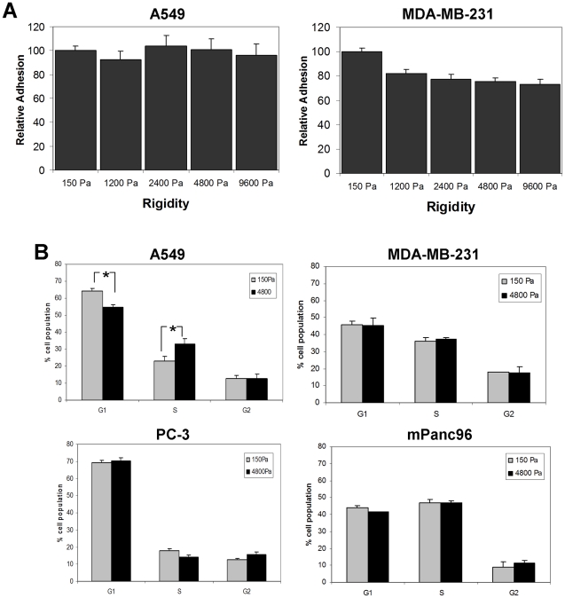
Background: The mechanical properties of the extracellular matrix have an important role in cell growth and differentiation. However, it is unclear as to what extent cancer cells respond to changes in the mechanical properties (rigidity/stiffness) of the microenvironment and how this response varies among cancer cell lines.

Methodology/principal findings: In this study we used a recently developed 96-well plate system that arrays extracellular matrix-conjugated polyacrylamide gels that increase in stiffness by at least 50-fold across the plate. This plate was used to determine how changes in the rigidity of the extracellular matrix modulate the biological properties of tumor cells. The cell lines tested fall into one of two categories based on their proliferation on substrates of differing stiffness: "rigidity dependent" (those which show an increase in cell growth as extracellular rigidity is increased), and "rigidity independent" (those which grow equally on both soft and stiff substrates). Cells which grew poorly on soft gels also showed decreased spreading and migration under these conditions. More importantly, seeding the cell lines into the lungs of nude mice revealed that the ability of cells to grow on soft gels in vitro correlated with their ability to grow in a soft tissue environment in vivo. The lung carcinoma line A549 responded to culture on soft gels by expressing the differentiated epithelial marker E-cadherin and decreasing the expression of the mesenchymal transcription factor Slug.

Conclusions/significance: These observations suggest that the mechanical properties of the matrix environment play a significant role in regulating the proliferation and the morphological properties of cancer cells. Further, the multiwell format of the soft-plate assay is a useful and effective adjunct to established 3-dimensional cell culture models.


Conflict of interest statement

Competing Interests: The authors have declared that no competing interests exist.

Figures

Figure 1
Figure 1. Design of the SoftPlate96 assay.
A typical 5-day growth assay using a soft-plate96 yields a “growth profile” which reflects the effect of rigidity on the proliferation of the cell line.
Figure 2
Figure 2. Soft-plate96 growth profiles of cancer cell lines.
The table is a compilation of 5-day growth assays for 17 cell lines. Included in the table are original source of the cells (indicated by literature citations), the ability to grow on 150 Pa and 9600 Pa substrates (from SoftPlate96 assays), and the soft-plate96 growth profile for each cell line. Grey profiles indicate rigidity-dependent lines and black profiles indicate rigidity-independent lines. Growth is measured as follows: −<1 fold; +1–5 fold; ++5–10 fold; +++ 10–15 fold; ++++ 15–20 fold; +++++ >20 fold increase in cell number over 5 days.
Figure 3
Figure 3. Growth of cancer cell lines on flexible substrates.
A.) 5-day growth assay of four cancer cell lines on plastic. B.) 5-day growth assays of the four cancer cell lines on a soft-plate96. Data are expressed as fold change over the number of cells initially plated. Results show mean ± SEM of at least three independent experiments. * p<0.05 vs. growth on 9600 Pa as measured by one-way ANOVA followed by Tukey's test.
Figure 4
Figure 4. Analysis of adhesion and cell cycle of cancer cell lines on soft gels.
A.) A549 and MDA-MB-231 cells were plated on a soft-plate96 and total cell numbers per well were counted after 6 hours of attachment. Data are expressed as percent of adhesion to the 150 Pa gels. B.) A549 and PC-3 cells were cultured on 150 Pa or 4800 Pa gel substrates for 5 days followed by cell cycle analysis. Results show average of at least three experiments ± SEM. * p<0.05.
Figure 5
Figure 5. Analysis of apoptosis of cancer cell lines on soft gels.
A.) Representative micrographs of A549 and MDA-MB-231 cells plated for 5 days on 150 Pa or 4800 Pa gel substrates. All cell nuclei are stained with DAPI (blue) and TUNEL-positive cells are labeled with fluorescein (green). Bar  = 100 µm. B.) Quantitation of TUNEL staining. Average of 2 experiments ± SEM with a total at least 400 cells counted for each condition.
Figure 6
Figure 6. The growth of cancer cell lines in mouse lung tissue.
A.) GFP-labeled MDA-MB-231, A549, PC-3, or mPanc96 cells were seeded into the lungs of nude mice. (Left) The number of GFP-positive cells in the lung was determined 4 hours and 14 days after injection, and the change in the number of GFP-positive cells in the lung over the 14 days was scored. * p<0.05 vs. MDA-MB-231 cells as measured by one-way ANOVA followed by Tukey's test. (Right) GFP-labeled A549 and mPanc96 cells were seeded into the lungs and the percentage of GFP-positive cells were scored at intervals over 24 hours. B.) Histology of the mouse lung at 14 days following injection of A549 cells (left panel) and mPanc96 cells (right panel). Arrows indicate micrometastases. C.) Comparison of the growth of cell lines on plastic (taken from Fig. 3A) and on 1200 Pa substrates (taken from Fig. 3B).
Figure 7
Figure 7. Rigidity-dependent changes in morphology and migration correlate with rigidity-dependent cell proliferation.
A.) Micrographs of A549, MDA-MB-231, PC-3, and mPanc96 cells that were plated on 150 or 4800 Pa gel substrates for 20 hours. B.) Areas of cells that were plated for 20 hours on 150 or 4800 Pa gel substrates. Results show mean fold increase over an unspread cell ± SEM of at least 20 cells counted for each condition. C.) A549, MDA-MB-231, PC-3, and mPanc96 cells were plated for 2 hours, then filmed for an additional 18 hours. Mean cell velocity ± SEM in µm/hr was determined by tracing and measuring the paths of 15 cells per rigidity per cell line. * p<0.05.
Figure 8
Figure 8. Substrate rigidity regulates E-cadherin expression in A549 cells.
A.) A549 cells were cultured for 3 days on gels with rigidities of 150, 4800, or 19200 Pa. Cells were fixed and stained for actin (green) and paxillin (red). Arrows indicate focal adhesions. B.) A549 cells were cultured on gels with rigidities of 150 or 19200 Pa. Cells were fixed and stained for actin (green) and E-cadherin (red). C.) A549 cells cultured on PA gels for 3 days were lysed and blotted for expression of E-cadherin, vimentin, and actin. D.) The relative levels of Slug and E-cadherin mRNA in A549 cells cultured on PA gels for 3 days as measured by real-time RT-PCR. Results show mean ± SEM of three independent experiments.

References

    1. Baum B, Settleman J, Quinlan MP. Transitions between epithelial and mesenchymal states in development and disease. Semin Cell Dev Biol. 2008;19:294–308. - PubMed
    1. Fre S, Vignjevic D, Schoumacher M, Duffy SL, Janssen KP, et al. Epithelial morphogenesis and intestinal cancer: new insights in signaling mechanisms. Adv Cancer Res. 2008;100:85–111. - PubMed
    1. Christofori G, Semb H. The role of the cell-adhesion molecule E-cadherin as a tumour-suppressor gene. Trends Biochem Sci. 1999;24:73–76. - PubMed
    1. Gagne D, Groulx JF, Benoit YD, Basora N, Herring E, et al. Integrin-linked kinase regulates migration and proliferation of human intestinal cells under a fibronectin-dependent mechanism. J Cell Physiol. 2010;222:387–400. - PMC - PubMed
    1. Moustakas A, Heldin CH. Signaling networks guiding epithelial-mesenchymal transitions during embryogenesis and cancer progression. Cancer Sci. 2007;98:1512–1520. - PMC - PubMed

Publication types