

doi: 10.1002/adma.201002448.
Collective cell migration on artificial extracellular matrix proteins containing full-length fibronectin domains
Affiliations
- PMID: 20886461
- PMCID: PMC3027490
- DOI: 10.1002/adma.201002448
Item in Clipboard
Collective cell migration on artificial extracellular matrix proteins containing full-length fibronectin domains
Adv Mater.
.
No abstract available
Figures
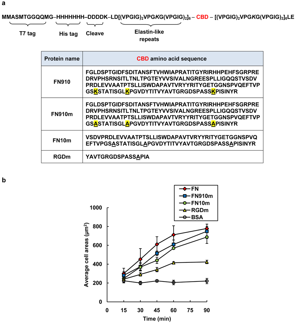
Artificial extracellular matrix (aECM) proteins containing full-length fibronectin domains. a) The general amino acid sequence of the aECM proteins. Each protein contains an N-terminal T7 tag, a hepahistidine tag, and an enterokinase cleavage site followed by six elastin-like repeats, a cell-binding domain (CBD, see table) and six elastin-like repeats. The differences between FN910 and FN910m are highlighted in yellow. The letter “m” denotes cell-binding domains containing lysine-to-alanine mutations. b) The average projected cell areas for the adsorbed protein surfaces at each time point. Data represent means ± SEM from three independent experiments.
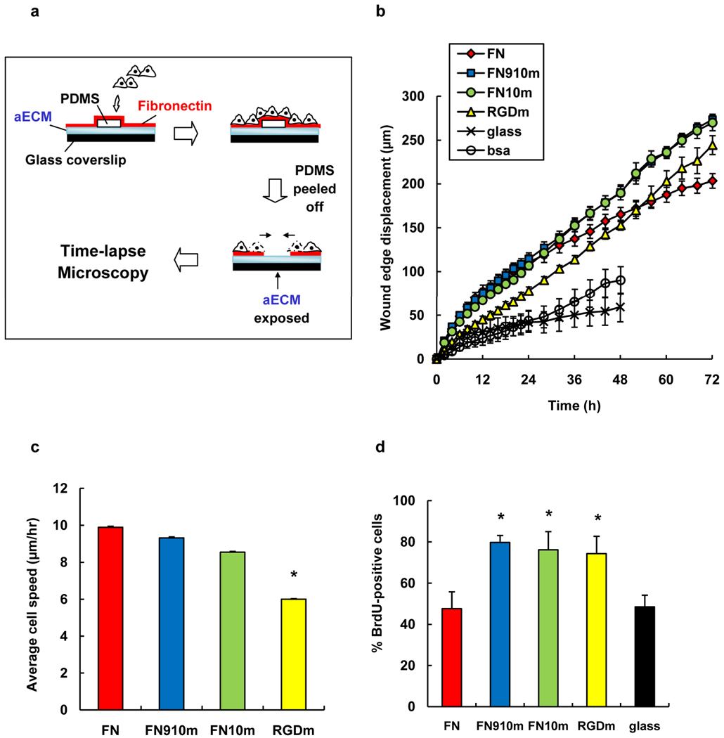
Wound-healing behavior of Rat-1 fibroblasts on adsorbed protein surfaces. a) Schematic of wound-healing assay. b) Displacement of the wound edge as a function of time. c) Average speeds of cells from t = 0 to 10 h. Individual cells in the first row of the wound edge were tracked for 10 h post wounding. Cell speeds are slopes from linear fit of average distance traveled vs. time. Error bars are standard errors from fit. d) The percentage of BrdU-positive cells for the period t = 24 to 48 h (the cell sheet was wounded at t = 0). Data are means ± SEM from five independent experiments for each surface. *, significant difference from FN surface (P < 0.05).
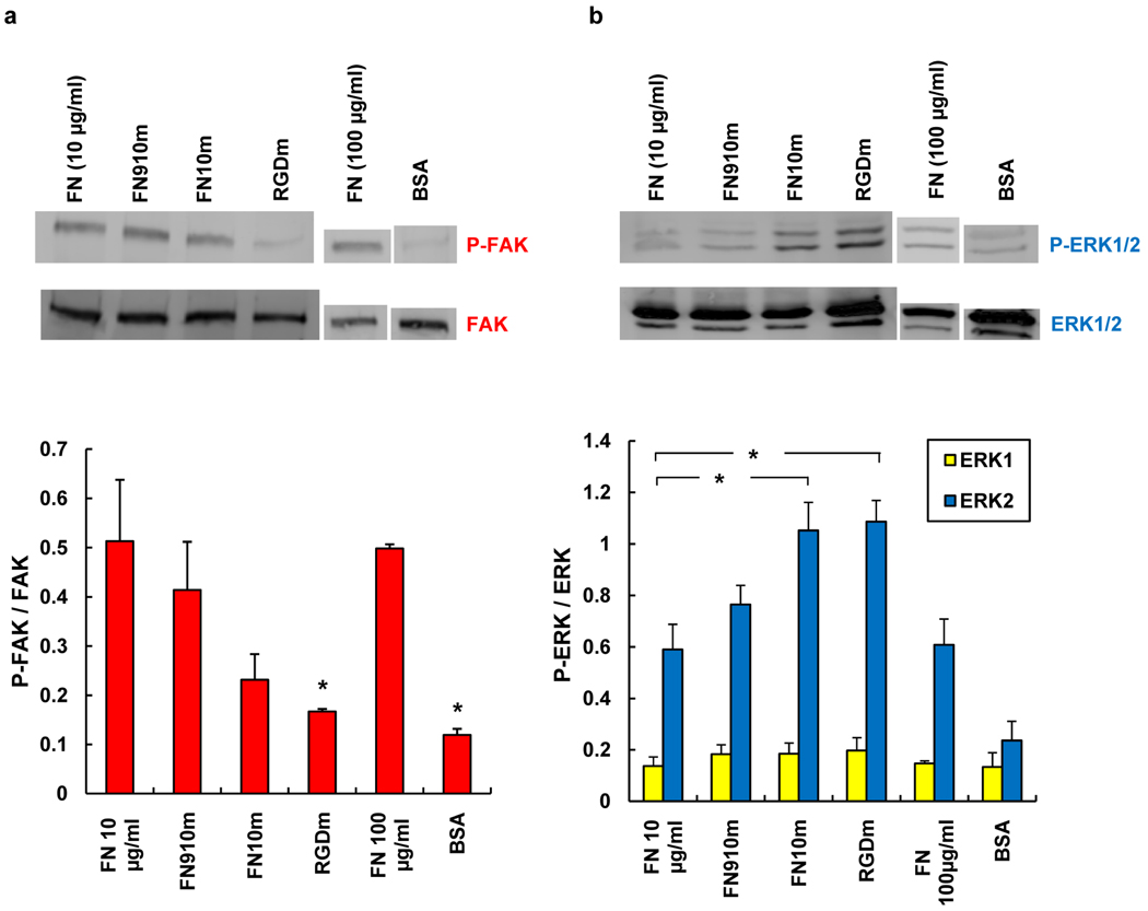
Determination of FAK and ERK phosphorylation in Rat-1 fibroblasts on adsorbed protein surfaces. Cell lysates were analyzed by Western blotting with a) anti-FAK, anti-phosphoFAK (pY397) and b) anti-phosphoERK1/2(p42/p44) and anti-total ERK1/2, antibodies. Band intensities were normalized to total-FAK or total-ERK bands. Reported data are means ± s.d. for three independent experiments. *, significantly different from FN surface (P < 0.05).
References
Publication types
MeSH terms
Substances
Grants and funding
LinkOut - more resources
Full Text Sources
