Review
. 2010:694:138-59.
doi: 10.1007/978-1-4419-7002-2_11.

Protein homeostasis in models of aging and age-related conformational disease

Affiliations
Review

Protein homeostasis in models of aging and age-related conformational disease

Elise A Kikis et al. Adv Exp Med Biol. 2010.

Abstract

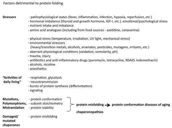
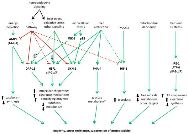
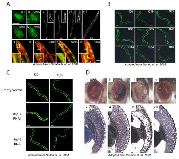
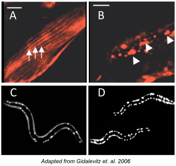
The stability of the proteome is crucial to the health of the cell, and contributes significantly to the lifespan of the organism. Aging and many age-related diseases have in common the expression of misfolded and damaged proteins. The chronic expression of damaged proteins during disease can have devastating consequences on protein homeostasis (proteostasis), resulting in disruption ofnumerous biological processes. This chapter discusses our current understanding of the various contributors to protein misfolding, and the mechanisms by which misfolding, and accompanied aggregation/toxicity, is accelerated by stress and aging. Invertebrate models have been instrumental in studying the processes related to aggregation and toxicity of disease-associated proteins and how dysregulation ofproteostasis leads to neurodegenerative diseases of aging.


Figures

Figure 1
Figure 1
Extrinsic and intrinsic factors detrimental to protein folding.
Figure 2
Figure 2. Proteostasis pathways
Multiple interconnected pathways regulate expression of genes that contribute to maintenance of protein folding homeostasis during growth, development, and under various stress conditions. The complex genetic interactions among these pathways necessitate precise control over their activities. Many of these pathways also possess regulatory feedback mechanisms, which are not well characterized/understood.
Figure 3
Figure 3
A) SOD1 mutant proteins aggregate in the body wall muscle cells of C. elegans. Fluorescent micrographs of SOD1transgenic embryos (i–iv) and adult animals (ix through xii) showing the distribution of the SOD-YFP fluorescence (green) and Rhodamine-phalloidin stained myofilaments (red). WT SOD1 protein exhibits diffuse, if patchy, fluorescence, while all mutant strains contain discrete fluorescent foci as well as some diffuse fluorescence. Panels v through viii show G85R animals in all larval stages (L1 through L4). B) Length-dependent aggregation of polyQ-YFP fusion proteins in C. elegans. Epifluoresence micrographs of 3- to 4-day-old C. elegans expressing different lengths of polyQ-YFP (Q0, Q19, Q29, Q33, Q35, Q40, Q44, Q64, Q82). C) RNAi for hsp-1 and hsf-1 induces aggregate formation of polyglutamine YFP proteins in a Q stretch length-dependent manner. Fluorescence microscopy pictures of 4-day-old C. elegans adults expressing Q0-YFP or Q35-YFP. RNAi of either hsp-1 or hsf-1 results in premature onset of aggregation in Q35 animals. D) HSP70 overexpression suppresses polyglutamine-induced neurodegeneration in Drosophila. Eyes (i–iv) and retinal sections (v,vi) of flies expressing expanded polyglutamine protein and human HSPA1L are shown. i,v, Control fly expressing only the promoter transgene. ii,vi, Flies expressing HSPA1L, coding for Hsp70 protein. Eye structure appears grossly normal. More detailed analysis revealed abnormalities in nuclear position and photoreceptor rhabdomere morphology when HSPA1L is highly expressed. iii,vii, Flies expressing the expanded polyglutamine protein MJDtr-Q78. These flies have degenerate eyes that lack pigment and show severe loss of retinal structure. iv,viii, Flies expressing both MJDtr-Q78 and HSPA1L. Co-expression of HSPA1L ameliorates the degenerative effects of MJDtr-Q78. The eye appears normal externally. Internally, eye structure is largely restored, although photoreceptor rhabdomere specializations are not made.
Figure 4
Figure 4. Progressive disruption of cellular folding capacity by misfolded proteins
(A and B) Confocal images showing cellular localization of ts mutant paramyosin in the absence (A) or presence (B) of Q40-YFP. Arrows, normal muscle sarcomeres, arrowheads, abnormal paramyosin(ts) assemblies in the presence of Q40, both under permissive conditions. (C and D) Images of Q40-YFP fluorescence in L2 larval stage animals expressing wild type (C) or ts mutant paramyosin (D) under permissive conditions.

References

    1. Rutherford SL, Lindquist S. Hsp90 as a capacitor for morphological evolution. Nature. 1998;396:336–342. - PubMed
    1. Stevens FJ, Argon Y. Pathogenic light chains and the B-cell repertoire. Immunol Today. 1999;20:451–457. - PubMed
    1. Gidalevitz T, Ben-Zvi A, Ho KH, Brignull HR, Morimoto RI. Progressive disruption of cellular protein folding in models of polyglutamine diseases. Science. 2006;311:1471–1474. - PubMed
    1. Yeyati PL, Bancewicz RM, Maule J, van Heyningen V. Hsp90 selectively modulates phenotype in vertebrate development. PLoS Genet. 2007;3:e43. - PMC - PubMed
    1. Michels AA, et al. Hsp70 and Hsp40 chaperone activities in the cytoplasm and the nucleus of mammalian cells. J Biol Chem. 1997;272:33283–33289. - PubMed

Publication types