Review
. 2011:6:19-48.
doi: 10.1146/annurev-pathol-011110-130327.

The pathogenesis of sepsis

Affiliations
Review

The pathogenesis of sepsis

Deborah J Stearns-Kurosawa et al. Annu Rev Pathol. 2011.

Abstract

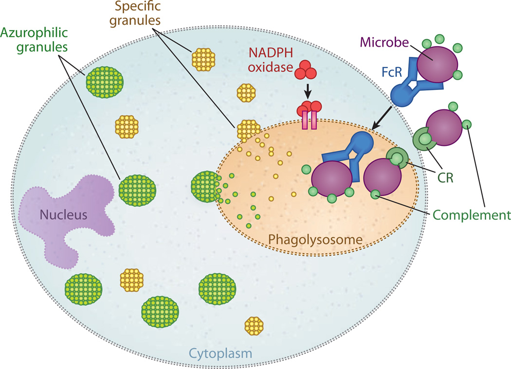
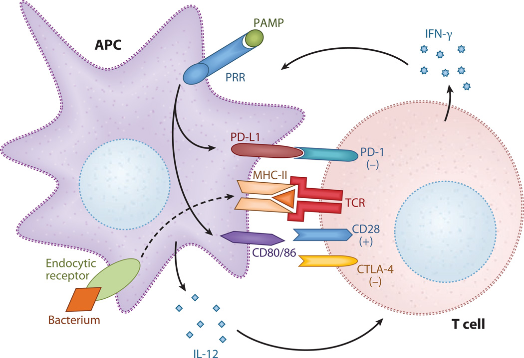
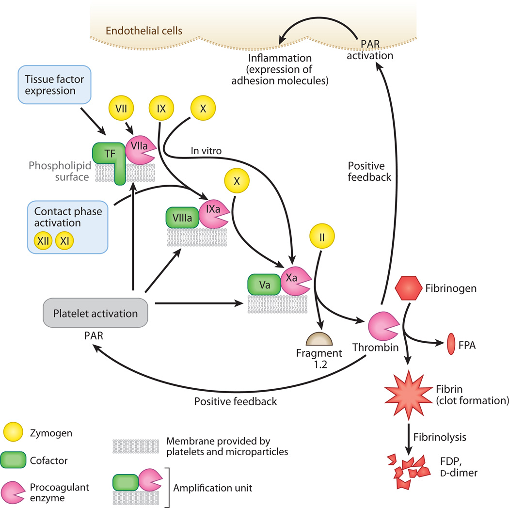
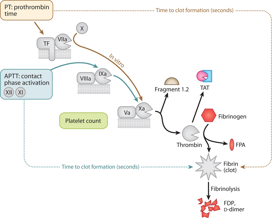
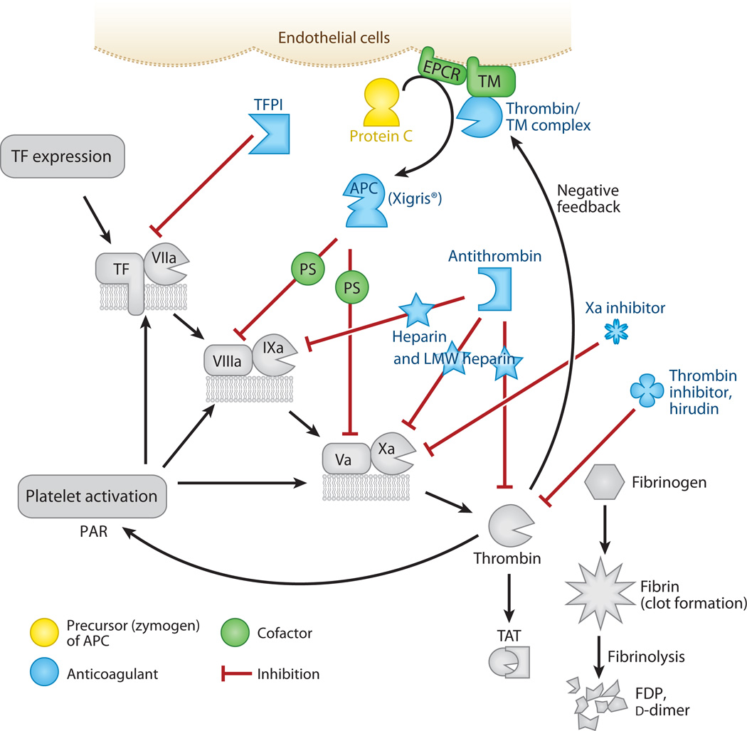
Sepsis is a serious clinical condition that represents a patient's response to a severe infection and has a very high mortality rate. Normal immune and physiologic responses eradicate pathogens, and the pathophysiology of sepsis is due to the inappropriate regulation of these normal reactions. In an ideal scenario, the first pathogen contact with the inflammatory system should eliminate the microbe and quickly return the host to homeostasis. The septic response may accelerate due to continued activation of neutrophils and macrophages/monocytes. Upregulation of lymphocyte costimulatory molecules and rapid lymphocyte apoptosis, delayed apoptosis of neutrophils, and enhanced necrosis of cells/tissues also contribute to the pathogenesis of sepsis. The coagulation system is closely tied to the inflammatory response, with cross talk between the two systems driving the dysregulated response. Biomarkers may be used to help diagnose patients with sepsis, and they may also help to identify patients who would benefit from immunomodulatory therapies.


Figures

Figure 1
Figure 1
Mechanisms of phagocytic-cell destruction of bacteria. Phagocytic cells such as neutrophils and macrophages are responsible for the clearance of pathogens from the body. Opsonization is the binding of antibodies and complement fragments to components on a bacterium. The antibodies bind to Fc receptors (FcRs) or complement receptors (CRs) on the surface of the phagocytic cell. After binding, the bacterium is taken into the cytoplasm; the membrane vacuole containing the phagocytosed bacteria is known as the phagosome. Fusion of the phagosome cytoplasmic granules such as specific granules (yellow) and azurophilic granules (green) forms the phagolysosome. Reactive oxygen intermediates are produced through the activity of NADPH oxidase, a complex of seven proteins. Adapted from Reference .
Figure 2
Figure 2
Costimulatory molecules (CSMs). Antigen presenting cells (APCs) detect infection through the binding of pathogen-associated molecular patterns (PAMPs) to pattern-recognition receptors (PRRs), as well as the phagocytosis of bacteria. Interleukin (IL)-12 is released, and expression of the CSMs (CD80, CD86, and PD-L1) is upregulated. These CSMs bind to corresponding T cell ligands, provided that the second signal as antigen is presented in the context of the major histocompatibility complex (MHC). CD80/86 binds to CD28, resulting in T cell activation and proliferation, and PD-L1:PD-1 interaction leads to T cell anergy and apoptosis. Ligation of cytotoxic T lymphocyte–associated antigen (CTLA)-4 expressed on previously activated T cells by CD80/86 provides a negative signal that regulates the degree of T cell activity. Interferon (IFN)-γ is released by the T cell–activating phagocytic cells to kill intracellular bacteria.
Figure 3
Figure 3
The formation of a clot. The coagulation cascade features a simple amplification unit that is repeated several times: A cofactor protein (green) binds to an enzyme (pink) on a phospholipid surface. This triumvirate complex is recognized by a circulating zymogen precursor (yellow). After a brief interaction, the complexed enzyme cleaves the zymogen and activates it. This new enzyme is incorporated into the next amplification unit for activation of the next precursor molecule. The tissue factor–Factor VIIa–membrane complex begins in vivo coagulation when tissue factor is exposed to blood components after injury. Factors XII and XI begin the contact phase–activation (intrinsic) pathway. Platelets provide the majority of phospholipid surfaces to support coagulation. Prothrombin (Factor II) is the precursor to thrombin, the final enzyme of the cascade, which releases Fragment 1.2 and thrombin during activation. Thrombin removes fibrinopeptides from fibrinogen, forming fibrin. When fibrin oligomerizes and is cross-linked, it provides surfaces for the fibrinolytic pathway, which comprises another set of activators and enzymes that cleave the clot and release fragments [fibrin degradation products (FDPs), d-dimers] into the circulation. Thrombin also acts in a proinflammatory manner by activating protease-activated receptors (PARs) on platelets and endothelial cells. Abbreviation: FPA, fibrinopeptide A.
Figure 4
Figure 4
Common tests in coagulation (colored entities). Steps discussed in previous figures are in gray. The prothrombin time (PT) is the number of seconds required to make a clot starting from the tissue factor (TF)–Factor VIIa–phospholipid unit. The activated partial thromboplastin time (APTT) is the number of seconds required to make a clot starting from the contact activation pathway. It is a partial clotting time because TF is not present. Both pathways merge at Factor Xa, which forms an amplification unit with Factor Va and lipid. Inhibitors of Factor Xa block the amplification unit and prevent thrombin formation. Tests are available to measure the presence of prothrombin Fragment 1.2 generated during thrombin production, the inactive thrombin-antithrombin complex (TAT), fibrinopeptide A (FPA) released during formation of fibrin, and chunks of cross-linked fibrin released during fibrinolysis of the clots [fibrin degradation products (FDPs), d-dimers].
Figure 5
Figure 5
The regulation of clot formation (colored entities). Steps discussed in previous figures are in gray. Regulation checkpoints during coagulation include tissue factor (TF), Factors VIIIa and Va, and Factor Xa. If these are inhibited, then thrombin production essentially stops. Tissue factor pathway inhibitor (TFPI) blocks the TF pathway. The protein C pathway blocks Factors VIIIa and Va. This pathway creates the activated protein C (APC) enzyme by the combined contributions of the endothelial protein C receptor (EPCR), thrombin, and thrombomodulin (TM) on cell surfaces. With the help of protein S (PS), APC cleaves the VIIIa and Va cofactors, which slows clotting by orders of magnitude. The drug Xigris® is recombinant human APC and is effective in patients with severe sepsis. Antithrombin is the natural inhibitor of several coagulation enzymes, including Factors IXa and Xa and thrombin. Its activity is accelerated by heparin and becomes specific for Factor Xa with low-molecular-weight (LMW) heparin. Hirudin is derived from leeches and inhibits only thrombin. Dysfunctional coagulation is common in sepsis patients, and the challenge is to prevent clot formation without increasing the risk of bleeding. Abbreviations: FDP, fibrin degradation product; PAR, protease-activated receptor; TAT, thrombin-antithrombin complex.

References

    1. Martin G, Brunkhorst FM, Janes JM, Reinhart K, Sundin DP, et al. The international PROGRESS registry of patients with severe sepsis: drotrecogin alfa (activated) use and patient outcomes. Crit. Care. 2009;13:R103. - PMC - PubMed
    1. Benoit M, Desnues B, Mege JL. Macrophage polarization in bacterial infections. J. Immunol. 2008;181:3733–3739. - PubMed
    1. Alcaide P, Auerbach S, Luscinskas FW. Neutrophil recruitment under shear flow: It’s all about endothelial cell rings and gaps. Microcirculation. 2009;16:43–57. - PMC - PubMed
    1. Urban CF, Lourido S, Zychlinsky A. How do microbes evade neutrophil killing? Cell Microbiol. 2006;8:1687–1696. - PubMed
    1. Le Tulzo Y, Pangault C, Gacouin A, Guilloux V, Tribut O, et al. Early circulating lymphocyte apoptosis in human septic shock is associated with poor outcome. Shock. 2002;18:487–494. - PubMed