. 2010 Oct;43(5):480-93.
doi: 10.1111/j.1365-2184.2010.00698.x.

Combination of photodynamic therapy with aspirin in human-derived lung adenocarcinoma cells affects proteasome activity and induces apoptosis

Affiliations

Combination of photodynamic therapy with aspirin in human-derived lung adenocarcinoma cells affects proteasome activity and induces apoptosis

A Chiaviello et al. Cell Prolif. 2010 Oct.

Abstract

Objectives: Photodynamic treatment (PDT) of human lung carcinoma cells A549 (p53(+/+)) and H1299 (p53(-/-)) induces fast but transient stalling of proteasome activity. We have explored the possibility of prolonging this effect by combining PDT with drugs capable of sustaining the stall, and promote apoptosis of surviving cells. We show that aspirin can be used to accomplish this.

Materials and methods: Cells were irradiated at doses ranging from 0.54 to 1.10 J cm(-2), and subsequently were incubated with aspirin at either high (10 and 5 mm) or low concentration (2.5 and 1.5 mm). Photofrin concentration and incubation time were constant (2.5 μg/ml and 16 h). Under these conditions, we analysed cell viability, colony-forming efficiency, cycle profile, expression patterns of specific proteins and ubiquitination state, after individual or combined administration.

Results: Treatment with either PDT or aspirin, rapidly induced proteasome malfunction and accumulation of cells in G(2)M, but did not induce apoptosis. However, when aspirin was added to cells (even at low concentrations) after PDT, the proteasome block was sustained. Moreover, significant cytotoxic effects, including apoptosis, were observed along with cytostatic effects (G(2)M accumulation/decreased colony formation).

Conclusions: Combination of PDT and low-toxicity drugs (such as aspirin) resulted in protracted inhibition of proteasome activity and induced apoptosis even in apoptosis-resistant cancer cells.


Figures

Figure 1
Figure 1
(a) Photodynamic therapy determines reversible arrest of proteasome activity. H1299 and A549 cells were incubated with 2.5 μg/ml Photofrin for 16 h then irradiated with 0.54 J cm−2 (low) or 1.10 J cm−2 (high) light fluences. Chymotrypsin‐like activity was measured at 2 and 6 h after photodynamic treatment. Columns represent mean of three independent experiments. Statistical analysis was performed using unpaired Student’s t‐test: *P < 0.01; **P < 0.001 and ***P < 0.0001. Values are expressed as percentage of basal (control) chymotrypsin‐like activity. (b) Sub‐lethal photodynamic treatment does not permanently inhibit proteasome function. Upper panels: Left and middle: H1299 and A549 cells were incubated with 2.5 μg/ml Photofrin for 16 h then irradiated (0.54 J cm−2). Right: H1299 cells were incubated with Photofrin as above and irradiated at double light fluence (1.10 J cm−2). Proteins were extracted from lysed cells 1, 2 and 3 h after PDT. Expression of BCL‐2, IκBα and p27KIP1 proteins was evaluated using western blotting. Nitrocellulose filters were reprobed with anti‐actin antibody to compare gel loads. Lower panels: Relative optical density changes were obtained by integrating electrophoretic bands from immunoblots. Data are expressed as relative changes (per cent of control). (c) Effect of dithiothreitol (DTT) on proteasome activity. Left panel: H1299 cells were treated with 1 mm DTT and irradiated as indicated. Cell proteins were extracted and analysed for expression of BCl‐2, p27KIP1 and IκBα at 1, 2, 3 and 6 h after PDT. Nitrocellulose filters were reprobed with anti‐actin (BCl‐2, p27KIP1) or anti‐tubulin (IκBα), to compare gel loads. Right panel: Reactive oxygen species formation was cytofluorimetrically evaluated in: (1) untreated H1299 cells (controls); (2) irradiated cells (0.54 J cm−2); and (3) irradiated cells (0.54 J cm−2) pre‐treated with 1 mm DTT.
Figure 2
Figure 2
(a) Photodynamic treatment promotes transient accumulation of ubiquitinated proteins. Ubiquitination electrophoretic profiles of proteins from H1299 cells incubated with Photofrin for 16 h but not irradiated (line 1); incubated in medium containing MG‐132 (10 μm) for 3 h (line 2); photodynamically treated (0.54 J cm−2) and extracted 1, 2 and 6 h after irradiation (lines 3, 4 and 5). Filter was reprobed with anti‐tubulin antibody to compare protein loads. (b) Photodynamic treatment affects half‐life of specific proteins. Left panel: H1299 cells incubated with 40 μg/ml of cycloheximide (CycloHex) for 0–3 h. After incubation, cells were washed and protein extracts were analysed for IκBα expression. Right panel: Cells incubated with cycloheximide for 3 h, washed and immediately subjected to PDT (0.54 J cm−2). Protein extracts were analysed for IκBα expression 1, 2 and 3 h after irradiation. Nitrocellulose filter was reprobed with anti‐tubulin antibody to compare gel loads. (c) Effect of a second PDT treatment. H1299 cells were irradiated, washed and incubated in a Photofrin‐free medium for 2 h. Then cells were subjected to a second photodynamic treatment at the same dose (0.54 J cm−2). Expression profile of three key proteins was evaluated by western blotting 1, 2 and 3 h after the second PDT treatment. Filters were reprobed with anti‐actin antibody to compare gel loads.
Figure 3
Figure 3
(a) Aspirin induces reversible arrest of proteasome activity. Upper panel: H1299 cells treated with 10 mm aspirin for 1, 2, 3 and 6 h. Expression profiles of BCL‐2 and p27KIP1 obtained by western blot analysis after protein extraction. Filter was reprobed with anti‐tubulin antibody to compare gel loads. Lower panel: Relative optical density changes obtained by integrating electrophoretic bands from immunoblots. Data expressed as relative changes (per cent of control). (b) Aspirin affects chymotrypsin‐like activity of H1299 cells in a time‐ and concentration‐dependent manner. Chymotrypsin‐like activity of untreated H1299 cells and cells incubated for 1, 3, 6 h with aspirin at three different concentrations (1.5, 2.5 and 10 mm). Experiment described in detail in Materials and methods section. Columns represent mean of three independent experiments. Statistical analysis was performed using unpaired Student’s t‐test: **P < 0.001 and ***P < 0.0001. (c, d) Influence of aspirin on viability of H1299 and A549 cells. Cells (H1299, panel c and A549, panel d) incubated for 24 or 48 h with 1.5, 2.5 and 10 mm aspirin. Cells were then released in aspirin‐free medium and analysed by MTS assay 24 h later. Columns represent mean of three independent experiments. Statistical analysis was performed using unpaired Student’s t‐test: **P < 0.001 and ***P < 0.0001.
Figure 4
Figure 4
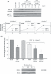
 Combined treatments: persistence of proteasome malfunction. (a) Upper panel: Effect of combined treatment on expression of BCL‐2 and p27KIP1 in H1299 and A549 cells. Extracts obtained from a) non‐irradiated H1299 and A549 cells (lines 1 and 2); (b) cells irradiated with a light dose of 0.54 J cm−2 and incubated with 5 or 10 mm aspirin for 24 h (lines 3 and 4 refer to H1299, lines 5 and 6, to A549); (c) H1299 cells irradiated with light dose of 1.10 J cm−2 and incubated with 1.5 or 2.5 mm aspirin for 72 h. Lower panel: Relative optical density changes obtained by integrating electrophoretic bands from immunoblots. Data expressed as relative changes (per cent of control). (b) Upper panel. Effect of combined treatment on chymotrypsin‐like activity in H1299 cells. From left: (a) untreated cells (control), (b) cells treated with 2.5 nm Bortezomib (used as positive control), (c) cells treated for 16 h with 2.5 μg/ml Photofrin (Ph) but not irradiated (dark effect), (d) cells irradiated using a fluence of 1.10 J cm−2 and (e) and (f) cells subjected to combined treatment (1.10 J cm−2 followed by 72 h treatment with 1.5 and 2.5 mm aspirin). Lower panel. Expression profile of BCL‐2 evaluated by western blotting after treatment with Bortezomib at 2.5 and 10 nm. Nitrocellulose filter was reprobed with anti‐actin antibody to compare gel load. Statistical analysis performed using unpaired Student’s t‐test: *P < 0.01; ***P < 0.0001.
Figure 5
Figure 5
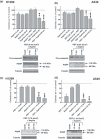
 Combined treatments: clonogenic survival and induction of apoptosis. Panels a and b: Effects of high aspirin concentration and low light fluence. Upper graphs: H1299 (left) and A549 (right) incubated for 24 h with 5 or 10 mm aspirin and then irradiated (aspirin + PDT), or first subjected to PDT then incubated with 5 or 10 mm aspirin for 24 h (PDT + aspirin). After treatments, cells were washed and incubated in fresh complete drug‐free medium. Colonies were stained with methylene blue ≥8 days later. Columns represent means of three independent experiments. Statistical analysis performed using unpaired Student’s t‐test: **P < 0.001; ***P < 0.0001. Colonies are expressed as per cent of untreated cells (dashed line). Lower panels: expression of pro‐caspase 3 and PARP in cells irradiated at 0.54 J cm−2 then incubated for 24 h with aspirin (5 and 10 mm). Nitrocellulose filter was reprobed with anti‐tubulin antibody to compare gel load. Panels c and d: Effects of low aspirin concentrations using higher light fluence. Upper graphs: H1299 (left) and A549 (right) cells irradiated with higher light fluence (1.10 J cm−2) washed and incubated for 72 h with 1.5 or 2.5 mm aspirin. Colonies stained with methylene blue ≥8 days later. Columns represent means of three independent experiments. Statistical analysis performed using unpaired Student’s t‐test: ***P < 0.0001. Colonies expressed as per cent of untreated cells (dashed line). Lower panels: expression of PARP in cells irradiated at 1.10 J cm−2 and then incubated for 24 h with aspirin (1.5 and 2.5 mm). Nitrocellulose filter reprobed with anti‐tubulin antibody to compare gel load.

Similar articles

Cited by

References

    1. Orlowski RZ (1999) The role of the ubiquitin‐proteasome pathway in apoptosis. Cell Death Differ. 6, 303–313. - PubMed
    1. Adams J (2004) The proteasome: a suitable antineoplastic target. Nat. Rev. Cancer 4, 349–360. - PubMed
    1. Karin M, Yamamoto Y, Wang QM (2004) The IKK NF‐kappaB system: a treasure trove for drug development. Nat. Rev. Drug Discov. 3, 17–26. - PubMed
    1. Fribley A, Zeng Q, Wang CY (2004) Proteasome inhibitor PS‐341 induces apoptosis through induction of endoplasmic reticulum stress‐reactive oxygen species in head and neck squamous cell carcinoma cells. Mol. Cell. Biol. 24, 9695–9704. - PMC - PubMed
    1. Hershko A (1997) Roles of ubiquitin‐mediated proteolysis in cell cycle control. Curr. Opin. Cell Biol. 9, 788–799. - PubMed

Publication types

MeSH terms