. 2010 Oct 1;143(1):35-45.
doi: 10.1016/j.cell.2010.09.004.

Myogenin and class II HDACs control neurogenic muscle atrophy by inducing E3 ubiquitin ligases

Affiliations

Myogenin and class II HDACs control neurogenic muscle atrophy by inducing E3 ubiquitin ligases

Viviana Moresi et al. Cell. .

Abstract

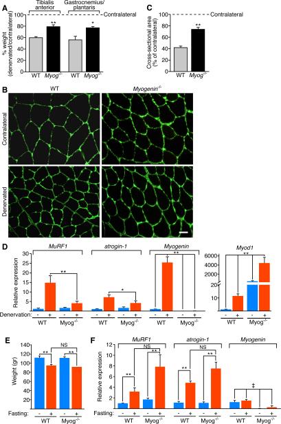
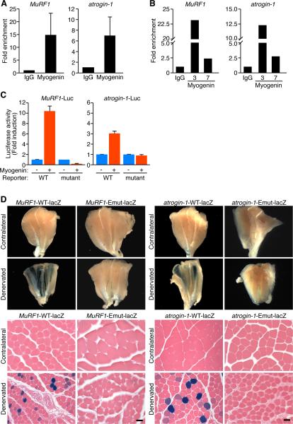
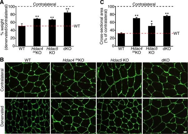
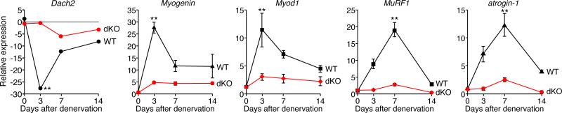
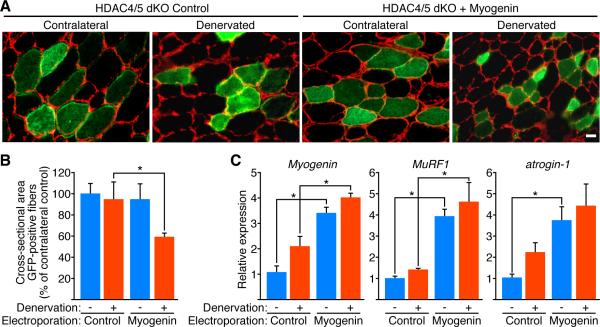
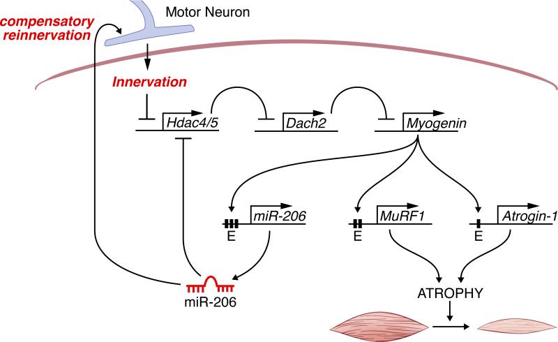
Maintenance of skeletal muscle structure and function requires innervation by motor neurons, such that denervation causes muscle atrophy. We show that myogenin, an essential regulator of muscle development, controls neurogenic atrophy. Myogenin is upregulated in skeletal muscle following denervation and regulates expression of the E3 ubiquitin ligases MuRF1 and atrogin-1, which promote muscle proteolysis and atrophy. Deletion of myogenin from adult mice diminishes expression of MuRF1 and atrogin-1 in denervated muscle and confers resistance to atrophy. Mice lacking histone deacetylases (HDACs) 4 and 5 in skeletal muscle fail to upregulate myogenin and also preserve muscle mass following denervation. Conversely, forced expression of myogenin in skeletal muscle of HDAC mutant mice restores muscle atrophy following denervation. Thus, myogenin plays a dual role as both a regulator of muscle development and an inducer of neurogenic atrophy. These findings reveal a specific pathway for muscle wasting and potential therapeutic targets for this disorder.


Figures

Figure 1
Figure 1. Adult Mice Lacking Myogenin Are Resistant to Muscle Atrophy Upon Denervation
(A) Percentage of TA or GP muscle weight of WT and Myog-/- mice 14 days after denervation, expressed relative to contralateral muscle. * p < 0.05 versus WT. ** p < 0.005 versus WT. n=4 for each sample. Data are represented as mean ± SEM. (B) Immunostaining for laminin of contralateral and denervated TA of WT and Myog-/- mice, 14 days after denervation. Scale bar=20 microns. (C) Morphometric analysis of contralateral and denervated TA of WT and Myog-/- mice, 14 days after denervation. Values indicate the mean of cross-sectional area of denervated TA fibers as a percentage of the contralateral fibers ± SEM. ** p < 0.005 versus WT. n=3 cross sections. (D) Expression of MuRF1, atrogin-1, Myogenin and Myod1 in contralateral (-) and denervated (+) GP of WT and Myog-/- mice, 7 days after denervation, detected by real-time PCR. The values are normalized to WT contralateral GP. Data are represented as mean ± SEM. * p < 0.05; ** p < 0.005 versus WT. n=4 for each sample. (E) Weight of GP muscle of WT and Myog-/- mice fed (-) or fasted (+) for 48 hours. Data are represented as mean ± SEM. ** p < 0.005 versus fed GP. NS=not significant. n=6 for each sample. (F) Expression of MuRF1, atrogin-1 and Myogenin in fed (-) and 48 hour fasted (+) GP of WT and Myog-/- mice, detected by real-time PCR. The values are normalized to WT fed GP. Data are represented as mean ± SEM. ‡ p < 0.005 versus WT. ** p < 0.005 versus fed. NS=not significant. n=6 for each sample. See also Figure S1 and S2.
Figure 2
Figure 2. Myogenin Directly Regulates MuRF1 and Atrogin-1
(A) ChIP assay performed in C2C12 myotubes showing myogenin binding to MuRF1 and atrogin-1 promoters. Chromatin was immunoprecipitated with antibodies against immunogloblulin G (IgG), or myogenin. Primers flanking the E-boxes on the MuRF1 and atrogin-1 promoters were used for amplifying DNA by real-time PCR. Values indicate the mean of fold enrichment over chromatin immunoprecipitated with antibodies against IgG ±SEM. n=3. (B) ChIP assays performed using denervated TA muscle at 3 and 7 days following denervation show myogenin binding to the MuRF1 and atrogin-1 promoters. Values indicate the fold enrichment over chromatin immunoprecipitated with antibodies against IgG. (C) Luciferase assays performed on cell extracts of C2C12 myoblasts transfected with luciferase reporter plasmids ligated to the WT (MuRF1-Luc) (atrogin-1-Luc), or the mutant constructs of MuRF1 and atrogin-1 genes, with myogenin (+) or empty (-) expression plasmid. Data are represented as mean ± SEM. (D) β-galactosidase staining of contralateral and denervated GP muscles isolated from transgenic mice containing a lacZ transgene under the control of the WT (MuRF1-WT-lacZ) (atrogin-1-WT-lacZ) or the mutant (MuRF1-Emut-lacZ) (atrogin-1-Emut-lacZ) constructs of the MuRF1 or atrogin-1 promoters. Upper panels show whole muscles. Lower panels show H&E sections. Scale bar=20 microns. See also Figure S3.
Figure 3
Figure 3. HDAC4 and HDAC5 Redundantly Regulate Skeletal Muscle Atrophy
(A) Percentage of TA muscle weight of mice of the indicated genotype 14 days after denervation, expressed relative to the contralateral muscle. Data are represented as mean ± SEM. ** p < 0.005 versus WT. n=5 for each sample. (B) Immunostaining for laminin in contralateral and denervated TA of mice of the indicated genotype, 14 days after denervation. Scale bar=20 microns. (C) Morphometric analysis of contralateral and denervated TA of indicated genotype, 14 days after denervation. Values indicate the mean of cross-sectional area of denervated TA fibers as a percentage of the contralateral fibers ± SEM. * p < 0.05 and ** p < 0.005 versus WT. n=3 cross sections. See also Figure S4 and S5.
Figure 4
Figure 4. dKO Mice Show Altered Gene Expression Upon Denervation
Expression of the indicated mRNAs was detected by real-time PCR in WT and dKO denervated GP and normalized to the expression in the contralateral muscle. Data are represented as mean ± SEM. ** p < 0.005 versus dKO. n=6 for each time point.
Figure 5
Figure 5. Ectopic Expression of Myogenin Induces Muscle Atrophy in dKO Mice Following Denervation
(A) Immunostaining for laminin (red) of cross-section of contralateral and denervated dKO TA electroporated with GFP expression plasmid and control plasmid (HDAC4/5 dKO Control) or GFP plasmid and myogenin (HDAC4/5 dKO + Myogenin), 10 days after denervation. Histology shows that the dKO denervated GFP-positive fibers co-electroporated with myogenin are smaller than denervated GFP-positive fibers co-electroporated with control plasmid. Scale bar=20 microns. (B) Morphometric analysis performed on GFP-positive fibers of contralateral (-) and denervated (+) dKO TA muscles electroporated with GFP expression plasmid and control plasmid (Control) or GFP plasmid and myogenin (Myogenin), 10 days after denervation. Values indicate the mean of cross-sectional area of GFP-positive muscle fibers as a percentage of the contralateral control fibers ± SEM. * p < 0.05 versus Control. n=7 for each condition. (C) Expression of Myogenin, MuRF1 and atrogin-1 in contralateral (-) and denervated (+) dKO TA muscles electroporated with GFP plasmid and a control plasmid (Control) or GFP plasmid and myogenin (Myogenin), 10 days after denervation. Values are normalized to the expression in the contralateral control muscles. Data are represented as mean ± SEM. * p < 0.05 versus Control. n=3 for each sample. See also Figure S6.
Figure 6
Figure 6. Model for Neurogenic Atrophy
Denervation of skeletal muscle results in the up-regulation of HDAC4 and HDAC5, which represses Dach2, a negative regulator of myogenin, resulting in myogenin expression. Myogenin activates the expression of MuRF1 and atrogin-1, two E3 ubiquitin ligases that participate in the proteolytic pathway resulting in muscle atrophy. Myogenin also regulates miR-206, which establishes a negative feedback loop to repress HDAC4 expression and promote reinnervation.

References

    1. Attaix D, Combaret L, Bechet D, Taillandier D. Role of the ubiquitin-proteasome pathway in muscle atrophy in cachexia. Curr. Opin. Support Palliat. Care. 2008;2:262–266. - PubMed
    1. Attaix D, Ventadour S, Codran A, Bechet D, Taillandier D, Combaret L. The ubiquitin-proteasome system and skeletal muscle wasting. Essays Biochem. 2005;41:173–186. - PubMed
    1. Backs J, Backs T, Bezprozvannaya S, McKinsey TA, Olson EN. Histone deacetylase 5 acquires calcium/calmodulin-dependent kinase II responsiveness by oligomerization with histone deacetylase 4. Mol. Cell. Biol. 2008;28:3437–3445. - PMC - PubMed
    1. Beehler BC, Sleph PG, Benmassaoud L, Grover GJ. Reduction of skeletal muscle atrophy by a proteasome inhibitor in a rat model of denervation. Exp. Biol. Med. 2006;231:335–341. - PubMed
    1. Blais A, Tsikitis M, Acosta-Alvear D, Sharan R, Kluger Y, Dynlacht BD. An initial blueprint for myogenic differentiation. Genes Dev. 2005;19:553–569. - PMC - PubMed

Publication types