. 2010 Nov 5;7(5):618-30.
doi: 10.1016/j.stem.2010.08.012. Epub 2010 Sep 30.

Highly efficient reprogramming to pluripotency and directed differentiation of human cells with synthetic modified mRNA

Affiliations

Highly efficient reprogramming to pluripotency and directed differentiation of human cells with synthetic modified mRNA

Luigi Warren et al. Cell Stem Cell. .

Abstract

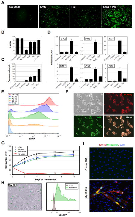
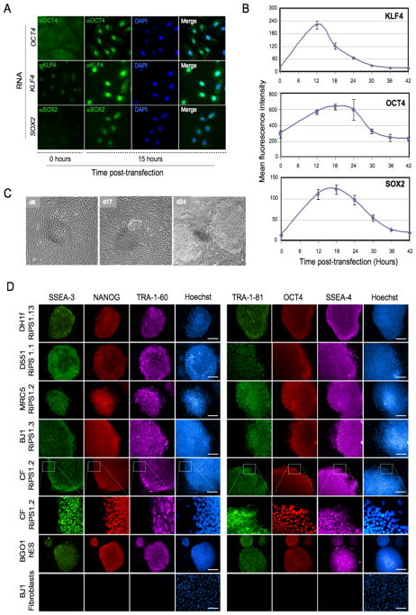
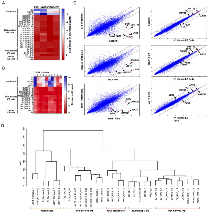
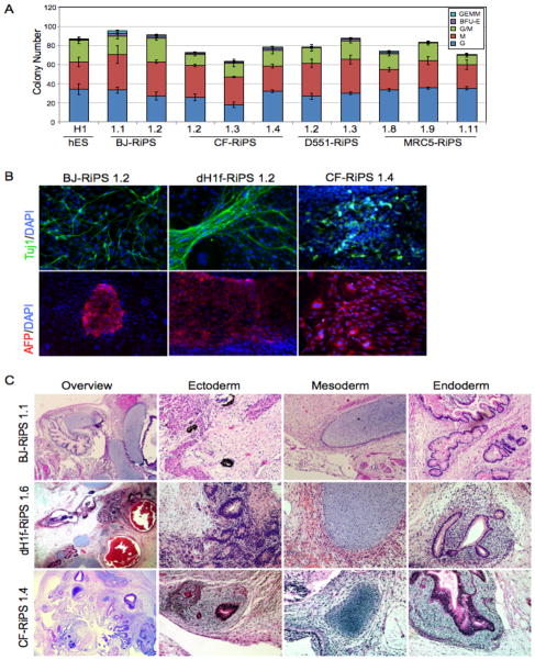
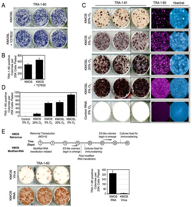
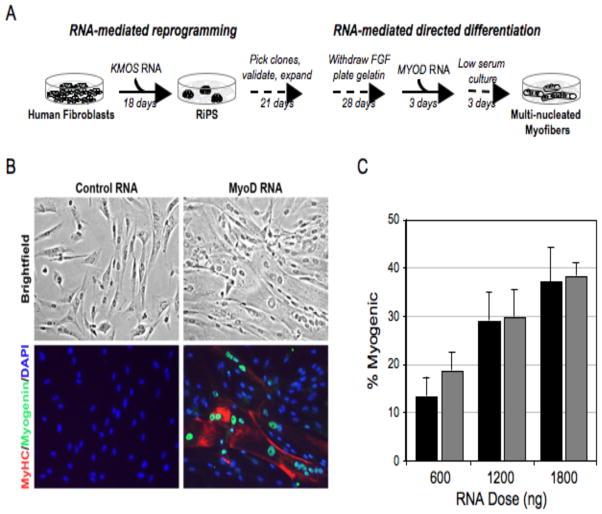
Clinical application of induced pluripotent stem cells (iPSCs) is limited by the low efficiency of iPSC derivation and the fact that most protocols modify the genome to effect cellular reprogramming. Moreover, safe and effective means of directing the fate of patient-specific iPSCs toward clinically useful cell types are lacking. Here we describe a simple, nonintegrating strategy for reprogramming cell fate based on administration of synthetic mRNA modified to overcome innate antiviral responses. We show that this approach can reprogram multiple human cell types to pluripotency with efficiencies that greatly surpass established protocols. We further show that the same technology can be used to efficiently direct the differentiation of RNA-induced pluripotent stem cells (RiPSCs) into terminally differentiated myogenic cells. This technology represents a safe, efficient strategy for somatic cell reprogramming and directing cell fate that has broad applicability for basic research, disease modeling, and regenerative medicine.


Figures

Figure 1
Figure 1. Modified-RNA overcomes anti-viral responses and can be used to direct cell fate
(A) Microscopy images showing keratinocytes transfected 24 hours earlier with 400 ng/well of synthetic unmodified (No Mods), 5-methyl-cytosine modified (5mC), pseudouridine modified (Psi), or 5mC + Psi modified-RNA encoding GFP. (B) Percent viability and (C) mean fluorescence intensity of the cells shown in (A) as measured by flow cytometry. (D) Quantitative RT-PCR data showing expression of six interferon-regulated genes in BJ fibroblasts 24 hours after transfection with unmodified (No Mods), or modified (5mC + Psi) RNA encoding GFP (1200 ng/well), and vehicle and untransfected controls. (E) Flow cytometry histograms showing GFP expression in keratinocytes transfected with 0–160 ng of modified-RNA, 24 hours post transfection. (F) Microscopy images of keratinocytes co-transfected with modified-RNAs encoding GFP with a nuclear localization signal, and mCherry (cytosolic) proteins. (G) Growth kinetics of BJ fibroblasts transfected daily with unmodified, or modified-RNAs encoding a destabilized nuclear-localized GFP, and vehicle and untransfected controls for 10 days. (H) Sustained GFP expression of modified-RNA transfected cells described in (G) at day 10 of transfection shown by fluorescence imaging with bright field overlay (left panel), and flow cytometry (right panel). (I) Immunostaining for the muscle-specific proteins myogenin and myosin heavy chain (MyHC) in murine C3H/10T1/2 cell cultures 3 days after 3 consecutive daily transfections with a modified-RNA encoding MYOD. Error bars indicate s.d., n=3 for all panels. See also Figure S2.
Figure 2
Figure 2. Generation of RNA-induced pluripotent stem cells (RiPS)
(A) Immunostaining for human KLF4, OCT4, and SOX2 proteins in keratinocytes 15 hours post-transfection with modified-RNA encoding KLF4, OCT4, or SOX2. (B) Time course showing kinetics and stability of KLF4, OCT4, and SOX2 proteins after modified-RNA transfection, assayed by flow cytometry following intracellular staining of reach protein. (C) Brightfield images taken during the derivation of RNA-iPS cells (RiPS) from dH1f fibroblasts showing early epitheliod morphology (day 6), small hES-like colonies (day 17), and appearance of mature iPS clones after mechanical picking and expansion (day 24). (D) Immunohistochemistry showing expression of a panel of pluripotency markers in expanded RiPS clones derived from dH1f fibroblasts, Detroit 551 (D551) and MRC-5 fetal fibroblasts, BJ post-natal fibroblasts, and cells derived from a skin biopsy taken from an adult cystic fibrosis patient (CF), shown also in high magnification. BG01 hES cells and BJ1 fibroblasts are included as positive and negative controls, respectively. See also Figure S3 and S4.
Figure 3
Figure 3. Molecular characterization of RiPS cells
(A) Heatmap showing results of qRT-PCR analysis measuring the expression of pluripotency-associated genes in RiPS cell lines, parental fibroblasts and viral-derived iPS cells relative to hES cell controls. (B) Heatmap showing results of OCT4 promoter methylation analysis of RiPS cell lines, parental fibroblasts, and hES cell controls. (C) Global gene expression profiles of BJ-, MRC5- and dH1F-derived RiPS cells shown in scatter plots against parental fibroblasts and hES cells with pluripotency-associated transcripts indicated. (D) Dendrogram showing unsupervised hierarchical clustering of the global expression profiles for RiPS cells, parental fibroblasts, hES cells, and virus-derived iPS cells.
Figure 4
Figure 4. Trilineage differentiation of RiPS cells
(A) Yield and typology of blood-lineage colonies produced by directed differentiation of embryoid bodies in methylcellulose assays with RiPS clones derived from BJ, CF, D551 and MCR5 fibroblasts, and a human ES (H1) control. (B) Immunostaining showing expression of the lineage markers Tuj1 (neuronal, ectodermal), and alpha-fetoprotein (epithelial, endodermal) in RiPS clones from 3 independent RiPS derivations subjected to directed differentiation. Hematoxylin and eosin staining of BJ-, CF- and dH1F-RiPS-derived teratomas showing histological overview, ectoderm (pigmented epithelia (BJ and CF), neural rosettes (dH1F)), mesoderm (cartilage, all), and endoderm (gut-like endothelium, all). For blood formation and methylcellulose assays, n=3 for each clone. See also Figure S5.
Figure 5
Figure 5. Pluripotency induction by modified-RNAs is highly efficient
(A) TRA-1-60 horseradish peroxidase (HRP) staining conducted at day 18 of a BJ-RiPS derivation with modified-RNAs encoding KMOSL and (B) frequency of TRA-1-60-positive colonies produced in the experiment relative to number of cells initially seeded. Error bars show s.d., n=6 for each condition. (C) TRA-181 HRP, TRA-160 immunofluorescence and Hoechst staining, and (D) colony frequencies for dH1f-RiPS experiments done using 4-factor (KMOS) and 5-factor (KMOSL) modified-RNA cocktails under 5% O2 or ambient oxygen culture conditions quantified at day 18. Control wells were transfected with equal doses of modified-RNA encoding GFP. (E) Kinetics and efficiency of retroviral and modified-RNA reprogramming. Timeline of colony formation (top panel), TRA-1-60 HRP immuno-staining (lower left panel), and TRA-1-60 positive colony counts (lower right panel) of dH1f cells reprogrammed using KMOS retroviruses (MOI=5 of each) or modified-RNA KMOS cocktails (n=3 for each condition). See also Figure S4.
Figure 6
Figure 6. Efficient directed differentiation of RiPS cells to terminally differentiated myogenic fate using modified-RNA
(A) Schematic of experimental design. (B) Bright-field and immunostained images showing large, multi-nucleated, myosin heavy chain (MyHC) and myogenin positive myotubes in cells fixed three days after cessation of MYOD modified-RNA transfection. Modified-RNA encoding GFP was administered to the controls. (C) Penetrance of myogenic conversion relative to daily RNA dose. Black bars refer to an experiment in which cultures were plated at 104 cells/cm2, grey bars to cultures plated at 5x10³ cells/cm2. Error bars show s.d. for triplicate wells.

Comment in

References

    1. Audouy S, Hoekstra D. Cationic lipid-mediated transfection in vitro and in vivo (review) Molecular membrane biology. 2001;18:129–143. - PubMed
    1. Chadwick K, Wang L, Li L, Menendez P, Murdoch B, Rouleau A, Bhatia M. Cytokines and BMP-4 promote hematopoietic differentiation of human embryonic stem cells. Blood. 2003;102:906–915. - PubMed
    1. Chan EM, Ratanasirintrawoot S, Park IH, Manos PD, Loh YH, Huo H, Miller JD, Hartung O, Rho J, Ince TA, et al. Live cell imaging distinguishes bona fide human iPS cells from partially reprogrammed cells. Nat Biotechnol. 2009;27:1033–1037. - PubMed
    1. Chang CW, Lai YS, Pawlik KM, Liu K, Sun CW, Li C, Schoeb TR, Townes TM. Polycistronic Lentiviral Vector for “Hit and Run” Reprogramming of Adult Skin Fibroblasts to Induced Pluripotent Stem Cells. Stem Cells. 2009;27:1042–1049. - PubMed
    1. Davis RL, Weintraub H, Lassar AB. Expression of a single transfected cDNA converts fibroblasts to myoblasts. Cell. 1987;51:987–1000. - PubMed

Publication types

MeSH terms

Associated data