. 2010 Nov 26;404(2):232-46.
doi: 10.1016/j.jmb.2010.09.034. Epub 2010 Oct 1.

Evolutionary origin of a secondary structure: π-helices as cryptic but widespread insertional variations of α-helices that enhance protein functionality

Affiliations

Evolutionary origin of a secondary structure: π-helices as cryptic but widespread insertional variations of α-helices that enhance protein functionality

Richard B Cooley et al. J Mol Biol. .

Abstract

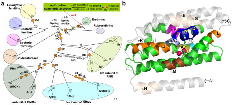
Formally annotated π-helices are rare in protein structures but have been correlated with functional sites. Here, we analyze protein structures to show that π-helices are the same as structures known as α-bulges, α-aneurisms, π-bulges, and looping outs, and are evolutionarily derived by the insertion of a single residue into an α-helix. This newly discovered evolutionary origin explains both why π-helices are cryptic, being rarely annotated despite occurring in 15% of known proteins, and why they tend to be associated with function. An analysis of π-helices in the diverse ferritin-like superfamily illustrates their tendency to be conserved in protein families and identifies a putative π-helix-containing primordial precursor, a "missing link" intermediary form of the ribonucleotide reductase family, vestigial π-helices, and a novel function for π-helices that we term a "peristaltic-like shift." This new understanding of π-helices paves the way for this generally overlooked motif to become a noteworthy feature that will aid in tracing the evolution of many protein families, guide investigations of protein and π-helix functionality, and contribute additional tools to the protein engineering toolkit.


Figures

Fig. 1
Fig. 1
The common two-H-bond π-helix is the same motif as an engineered α-aneurism. a) An α-helix in Staphylococcus aureus nuclease (left, green cartoon; PDB 1eyd) develops an αaneurism (right, purple cartoon; PDB 1sty) when a single amino acid (highlighted blue) is inserted into it. b) An overlay of the π-helix from fumarase C of Escherichia coli (residues 155–161, orange carbon atoms; PDB 1fur) and the same engineered α-aneurism from panel a (purple carbon atoms) demonstrates their equivalence. These α-aneurism-type helical distortions have been independently characterized as looping outs, π-bulges and α-bulges, but not as πhelices. Black-dashed lines represent the π-type H-bonds. (φ,Ψ) torsion angles are written beside each residue. In panel b, nitrogen (blue) and oxygen (red) atoms are indicated.
Fig. 2
Fig. 2
Defining π-helices based on (i+5,i) π-type H-bonds. a) Energy cutoffs (in kcal/mol, calculated by DSSP17) for strong (s), medium (m), and weak (w) H-bonds. b) Excerpted DSSP output for residues 245–267 of methane monooxygenase hydroxylase (MMOH) (PDB 1mty, βchain), highlighting the π-type H-bonding patterns. Green boxes highlight π-type donor interactions and red boxes highlight the acceptor interactions. Solid outlines indicate the π-type interaction is the strongest H-bond for that residue, and dotted outlines indicate those that are the second strongest. The column contents are labeled. For each of the four H-bond interaction columns, the two numbers given are relative positions of the partner residue and the energy of the interaction (in kcal/mol). The first two of these columns are the strongest interactions and the second two columns are the second strongest interactions. c) Short hand summary of the πtype H-bonding and the π-helix assignments. For each residue acting as a π-type donor or acceptor, the strength of the interaction is given in the appropriate column. The letter is in parentheses if the π-type interaction is not the strongest H-bond for that residue. Based on the qualifying criteria described in the Materials and Methods, this segment in MMOH that was previously classified as a single 20 residue π-helix with 5-(i+5,i) H-bonds is seen to be three distinct, but overlapping π-helices with 2-, 2- and 4-H-bonds (see rectangles in panel c). Note that these three π-helices are cryptic in that none are assigned as π-helical (I) in the DSSP summary (fourth column).
Fig. 3
Fig. 3
A single insertion can generate all occurring lengths of π-helices. Panels a, b, c, d, e and f show one example each of a 2-, 3-, 4-, 5-, 6- and 7-H-bond π-helix, respectively. In each panel, the equivalent α-helix (left) and π-helix (right) from a pair of homologous proteins are shown with their PDB codes. Above the black arrow that represents the insertion process is the sequence alignment from the FSSP database illustrating the single insertion. The π-helices from panels a (formaldehyde ferredoxin oxidoreductase), b (glycogen phosphorylase), and d (MMOH α-subunit; insertion πD from Fig. 7) are all located within the active site of their respective protein, while that shown in panel c (MMOH α-subunit) and panel e (α-subunit of Toluene-4-Monooxygenase; insertion πE from Fig. 7) are involved in intersubunit interactions, suggesting a functional role for these. All atoms in the α-helices are colored green, while carbon, nitrogen and oxygen atoms in the π-helices are colored orange, blue and red, respectively. Black dashed lines in the π-helices indicate the (i+5,i) π-type hydrogen bonds.
Fig. 4
Fig. 4
Evidence that overlapping π-helices result from stepwise insertions of single amino acids. In the ferritin-like superfamily, putative ancestral forms in the evolution of the three, overlapping π-helices in the β-subunit of MMOH (shown in Fig. 2) (bottom) are represented by the structurally equivalent helices from bacterioferritin (top), which is a pure α-helix, the R2 subunit of class Ib ribonucleotide reductase (second from top), which includes one π-helix, and the R2 subunit of class Ic ribonucleotide reductase (third from top), which includes overlapping πhelices. Insertion events represented are designated πB, πD and πJ in Figure 7. Amino acid alignments indicate the effective points of insertion. Lines below each sequence indicate the πhelical regions. π-helices are colored orange, α-helices are colored green, and the inserted amino acid, as defined by structural alignment, is colored in blue.
Fig. 5
Fig. 5
Three examples of α-helix derived active site π-helices. a) Evolutionary path by which π-helices are derived from α-helices via a single residue insertion (orange circle). b) Shown is the π-helix-containing PLP-bound active site of PLP-dependent glycogen phosphorylase (orange carbon atoms; PDB 3gpb) overlaid onto the α-helix-containing active site of trehalose-6-phosphate synthase (semitransparent green carbon atoms; PDB 1uqu). The structurally derived sequence alignments are shown below the structure (orange and green letters correspond to the π- and α-helix-containing sequences, respectively), which demonstrate the presence of the inserted residue that forms the π-helical segment (orange bar). c) The active site π-helix of mercuric ion reductase (orange carbon atoms; PDB 1zk7) is overlaid on the homologous α-helix of dihydrolipoamide dehydrogenase (semitransparent green carbon atoms; PDB 1ebd). d) A πhelix of acetylcholine esterase (orange carbon atoms; PDB 1ea5) is overlaid onto the homologous α-helix of mycolyl transferase (green transparent carbon atoms; PDB 1f0p). In all panels, dashed-black lines show the π-type H-bonds and red dashed lines show interactions within the active sites. Nitrogen (blue), oxygen (red), sulfur (yellow) and phosphorus (purple) atoms are indicated.
Fig. 6
Fig. 6
π-helical peristalsis in the active site of BMM enzymes. A) Two π-helices (labeled πB and πD) in the active site of MMOH are overlapping in the resting state (left). Upon binding the product analogue 6-bromohexan-1-ol (blue spheres), these two π-helices no longer overlap due to a shift in πB (right). B) The resting state structure of the toluene-4-monooxygenase hydroxylase also shows two overlapping π-helices at the active site (left). Upon binding of the regulatory component (ToMOD, semitransparent yellow), πB shifts identically to that of product-bound MMOH. As πB moves downward, an adjacent π-helix (labeled πE), which is sandwiched between the active site π-helix and the regulatory subunit, simultaneously elongates and shifts upward. For details of the π-type H-bonds, see Supplemental Figure 3. Orange bars mark individual π-helices and blue arrows the direction of π-helix movement. π-type H-bonds are shown by black dashed lines. π-helix (orange) and α-helix carbon atoms are indicated, as are nitrogen (blue), oxygen (red), iron (purple) and bromine (brown). PDB codes are 1mty, 1xvb, 3dhg and 3dhi.
Fig. 7
Fig. 7
π-helices and the evolution of the ferritin-like superfamily. a) Evolutionary tree showing that, from a symmetrical erythrin-like ancestor containing two π-helices (Fig. 8), a simple set of insertion (orange circles) and deletion (crossed-out orange circles) events can account for all πhelices seen in modern-day family members (Supplemental Table 5). The branching shown is based on maximum likelihood analysis as previously described from structurally-derived sequence alignments (bootstrap values shown). Organism names, omitted for clarity, are shown in Supplemental Figure 4. The length of the bar (bottom right) corresponds to 0.5 substitutions/site. New abbreviations are PH: phenol hydroxylase family and ArH: aromatic monooxygenase hydroxylase family. b) Ribbon diagram of the α-subunit of MMOH (PDB 1mty, chain D) showing the locations of its six π-helices (πB, πD, πE-πH) and also showing the locations of the remaining eight π-helices seen in other members of the ferritin-like superfamily (πA1, πA2, πC, πI-πM). Indicated are the core four-helix bundle (green), the surrounding helices (transparent gray), and the π-helices πA1 (yellow), πA2 (light blue), πC (cyan), πB and πD (blue), πE and πG-πL (orange), πF (purple) and πM (brown). Iron atoms (pink spheres) and the N- and C-termini are shown.
Fig. 8
Fig. 8
Model for the origins of the ferritin-like superfamily. a) Scheme showing the formation of the four-helix bundle characteristic of the ferritin-like superfamily via gene duplication and fusion of a primordial two-helix precursor that, through a single insertion πA (orange circle), gave rise to two symmetry related π-helices (πA1 and πA2) and eight ligating residue in the erythrin-like precursor of this superfamily. b) Sequence alignments showing the residual internal sequence similarity in erythrins and, to a lesser degree, rubrerythrin. Shown are the alignments of helix A with helix C and helix B with helix D in erythrin from Cyanophora paradoxa (CP Ery), erythrin from Gloeobacter violaceus (GV Ery), rubrerythrin from Desulfovibrio vulgaris (Rub) and bacterioferritin from Escherichia coli (Bfr) included as an example of a canonical superfamily member having only six ligating residues and less recognized internal symmetry. Red highlighting indicates residues involved (or proposed to be involved in the case of erythrin) in iron-ligation. Green highlighting indicates additional residues that are identical between homologous helices within the same protein. Orange bars indicate π-helical residues in rubrerythrin, and potential π-helical residues in erythrin.
Fig. 9
Fig. 9
π-helices πA2- πD correlate with changes in metallocenter geometry. Shown is an annotated view of the di-nuclear site in RNR Ic (PDB 1syy) with the backbone ribbon of core-helix B of the four-helix bundle, contributing H123 and E120, omitted for clarity. Core helices A, C and D and their direction from N- to C-terminus are labeled on the left, and π- (orange) and α-helices (green) are indicated. As described in the text, the proximity of πA2-πD to their respective di-nuclear centers are shown and suggest they play a role in influencing metallocenter chemistry.

References

    1. Bloom JD, Arnold FH. In the light of directed evolution: pathways of adaptive protein evolution. Proc Natl Acad Sci U S A. 2009;106(Suppl 1):9995–10000. - PMC - PubMed
    1. Dean AM, Thornton JW. Mechanistic approaches to the study of evolution: the functional synthesis. Nat Rev Genet. 2007;8:675–88. - PMC - PubMed
    1. Worth CL, Gong S, Blundell TL. Structural and functional constraints in the evolution of protein families. Nat Rev Mol Cell Biol. 2009;10:709–20. - PubMed
    1. Low BW, Baybutt RB. The π-helix- A Hydrogen Bonded Configuration of the Polypeptide Chain. J Am Chem Soc. 1952;74:5806–5807.
    1. Hollingsworth SA, Berkholz DS, Karplus PA. On the occurrence of linear groups in proteins. Protein Sci. 2009;18:1321–5. - PMC - PubMed

Publication types