. 2010:483:1-29.
doi: 10.1016/S0076-6879(10)83001-0.

Analyses of subnanometer resolution cryo-EM density maps

Affiliations

Analyses of subnanometer resolution cryo-EM density maps

Matthew L Baker et al. Methods Enzymol. 2010.

Abstract

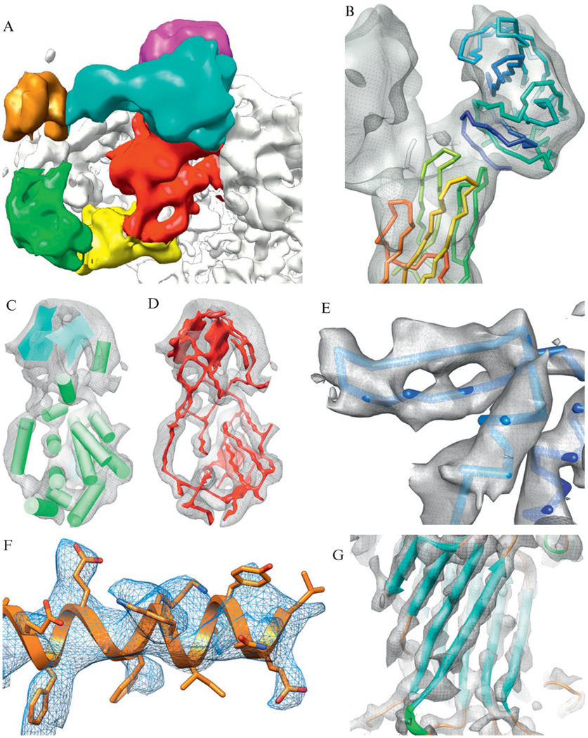
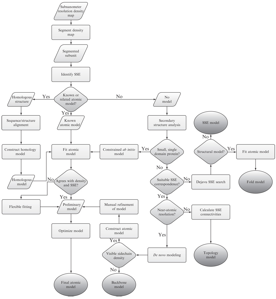
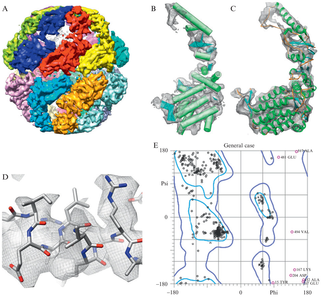
Today, electron cryomicroscopy (cryo-EM) can routinely achieve subnanometer resolutions of complex macromolecular assemblies. From a density map, one can extract key structural and functional information using a variety of computational analysis tools. At subnanometer resolution, these tools make it possible to isolate individual subunits, identify secondary structures, and accurately fit atomic models. With several cryo-EM studies achieving resolutions beyond 5Å, computational modeling and feature recognition tools have been employed to construct backbone and atomic models of the protein components directly from a density map. In this chapter, we describe several common classes of computational tools that can be used to analyze and model subnanometer resolution reconstructions from cryo-EM. A general protocol for analyzing subnanometer resolution density maps is presented along with a full description of steps used in analyzing the 4.3Å resolution structure of Mm-cpn.


Figures

Figure 1.1
Figure 1.1
Features at subnanometer resolutions. A gallery of structural features from cryo-EM reconstructions is shown. (A) Domains in the clamp region of the 9.5 Å resolution reconstruction of RyR1 can be observed (Serysheva et al., 2008). (B) The atomic models of VP5* (lower left) and VP8* (upper right) are fit to the density map corresponding to the VP4 spikes in the 9.5 Å resolution structure of rotavirus (Li et al., 2009). (C) At slightly higher resolutions, secondary structures (α-helices are depicted as cylinders and β-sheets are depicted as planes) can be clearly seen in the capsid proteins of rice dwarf virus at 6.8 Å resolution (Zhou et al., 2001). (D) Around this resolution, possible connections between secondary structure elements can be identified computationally using density skeletonization (red), again as seen in the 6.8 Å resolution rice dwarf virus capsid protein. (E) Increasing resolution reveals the separation of β-strands in GroEL at 4.2 Å resolution (Ludtke et al., 2008). (F) Large, bulky sidechains begin to appear in TriC reconstruction at 4.0 Å resolution (Cong et al., 2010). (G) An unambiguous backbone is apparent in VP6 of rotavirus at ~3.8 Å resolution (Zhang et al., 2008).
Figure 1.2
Figure 1.2
Analyzing a subnanometer resolution cryo-EM density map. A general scheme for analyzing subnanometer resolution cryo-EM density maps is depicted. Different projects may take advantage of additional information during the analysis process and thus deviate from the overall scheme.
Figure 1.3
Figure 1.3
Secondary structure identification. SSEHunter and SSEBuilder, both EMAN programs, can be run as plug-ins to UCSF’s Chimera. The results for the apical domain of Mm-cpn are shown: red spheres represent helix like regions and the blue spheres represent sheet like regions. Regions of similar scoring pseudoatoms from SSEHunter are grouped and built using SSEBuilder. Helices are depicted as cylinders and sheets are depicted as planes.
Figure 1.4
Figure 1.4
Model construction with Gorgon. The results from the SSE correspondence search on the apical domain of the 4.2-Å resolution structure of GroEL in Gorgon are shown in (A). Helices are shown as cylinders, while sheets are shown as planes. Potential connectivity is depicted as solid lines. A corresponding color scheme for these elements is shown in the SSE correspondence window on the right. (B) Gorgon contains several methods for assigning atoms to the density in the semi-automated atom placement tool. The atomic editor function illustrates the addition of Cα atoms along a density skeleton (not shown). The user can cycle through the possible locations, select the desired position and proceed to the next residue.
Figure 1.5
Figure 1.5
Model optimization and validation. Coot can be used to adjust mainchain and sidechain atom positions, optimizing the fit of the atomic model in the density map. A Ramachandran plot is shown overlaid with the map and model of Mm-cpn after optimization.
Figure 1.6
Figure 1.6
Structure of Mm-cpn. (A) The 4.3-Å resolution structure of Mm-cpn is shown (Zhang et al., 2010). (B) Using SSEHunter, the secondary structure elements in the Mm-cpn subunit were identified: α-helices are shown as cylinders and β-strands are shown as planes. (C) Using the de novo modeling approach, an atomic model (residues 1–532) was constructed for one subunit of Mm-cpn. (D) Large, bulky sidechains in the model could be seen in the density. (E) The Ramachandran plot of an Mm-cpn monomer shows greater than 98% of all residues with allowable phi–psi angles.

References

    1. Abeysinghe S, Ju T, Baker ML, Chiu W. Shape modeling and matching in identifying 3D protein structures. Comput. Aided Des. 2008;40:708–720.
    1. Altschul SF, Gish W, Miller W, Myers EW, Lipman DJ. Basic local alignment search tool. J. Mol. Biol. 1990;215:403–410. - PubMed
    1. Arnold K, Bordoli L, Kopp J, Schwede T. The SWISS-MODEL workspace: A web-based environment for protein structure homology modelling. Bioinformatics. 2006;22:195–201. - PubMed
    1. Baker ML, Jiang W, Bowman BR, Zhou ZH, Quiocho FA, Rixon FJ, Chiu W. Architecture of the herpes simplex virus major capsid protein derived from structural bioinformatics. J. Mol. Biol. 2003;331:447–456. - PubMed
    1. Baker ML, Jiang W, Rixon FJ, Chiu W. Common ancestry of herpesviruses and tailed DNA bacteriophages. J. Virol. 2005;79:14967–14970. - PMC - PubMed

Publication types

MeSH terms

Substances

LinkOut - more resources