Review
. 2011 Apr;1813(4):616-22.
doi: 10.1016/j.bbamcr.2010.09.013. Epub 2010 Oct 1.

Is mPTP the gatekeeper for necrosis, apoptosis, or both?

Affiliations
Review

Is mPTP the gatekeeper for necrosis, apoptosis, or both?

Kathleen W Kinnally et al. Biochim Biophys Acta. 2011 Apr.

Abstract

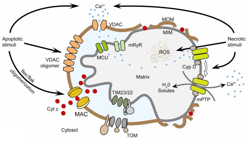
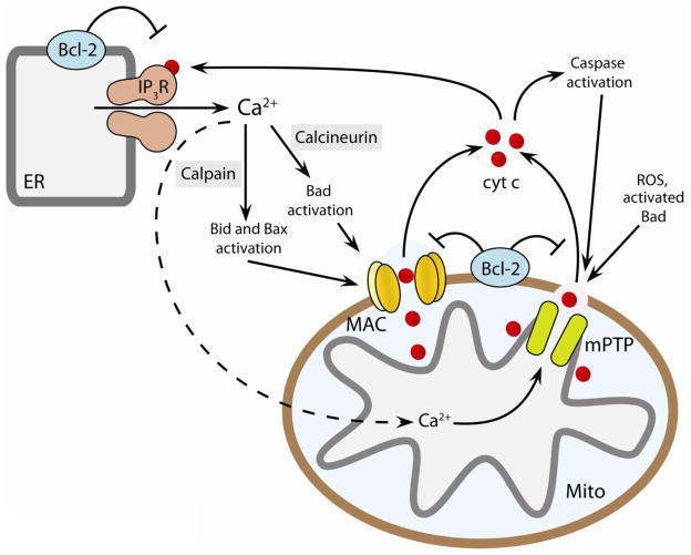
Permeabilization of the mitochondrial membranes is a crucial step in apoptosis and necrosis. This phenomenon allows the release of mitochondrial death factors, which trigger or facilitate different signaling cascades ultimately causing the execution of the cell. The mitochondrial permeability transition pore (mPTP) has long been known as one of the main regulators of mitochondria during cell death. mPTP opening can lead to matrix swelling, subsequent rupture of the outer membrane, and a nonspecific release of intermembrane space proteins into the cytosol. While mPTP was purportedly associated with early apoptosis, recent observations suggest that mitochondrial permeabilization mediated by mPTP is generally more closely linked to events of late apoptosis and necrosis. Mechanisms of mitochondrial membrane permeabilization during cell death, involving three different mitochondrial channels, have been postulated. These include the mPTP in the inner membrane, and the mitochondrial apoptosis-induced channel (MAC) and voltage-dependent anion-selective channel (VDAC) in the outer membrane. New developments on mPTP structure and function, and the involvement of mPTP, MAC, and VDAC in permeabilization of mitochondrial membranes during cell death are explored. This article is part of a Special Issue entitled Mitochondria: the deadly organelle.


Figures

Figure 1
Figure 1. Mitochondrial ion channels in apoptosis and necrosis
Left, Apoptotic stimuli induce relocation of Bax from the cytosol into the mitochondrial outer membrane (MOM). Bax, Bak, and possibly other unidentified protein(s) oligomerize and form MAC to release cytochrome c. VDAC oligomerization upon apoptotic stimuli was also reported to be involved in cytochrome c release. Right, Necrotic stimuli lead to exacerbated calcium uptake and reactive oxygen species generation by mitochondria. High levels of calcium and reactive oxygen species (ROS) induce a cyclophilin-D (Cyp D)-sensitive opening of mPTP that leads to swelling of the matrix and release of calcium. Swelling disrupts the outer membrane while released calcium activates proteases, phosphatases and nucleases that lead to necrotic degradation. Adapted from [112]. TIM23/22, translocase of the inner membrane complexes 23/22; mRyR, mitochondrial ryanodine receptor; TOM, translocase of the outer membrane; MCU, mitochondrial calcium uniporter; MIM, mitochondrial inner membrane.
Figure 2
Figure 2. The channel activities of VDAC, mPTP and MAC
Current traces were recorded from patches excised from either reconstituted outer (MAC and VDAC) or native inner membranes (mPTP) under symmetrical 150 mM KCl. Current traces are represented in the same scale for comparison. Current levels of open (O) channels are shown with downward transitions to closed (C) states in all the traces. Mouse MAC and VDAC traces were adapted from ref. [62], while mouse mPTP was from ref. [56].
Figure 3
Figure 3. Crosstalk between MAC and mPTP amplifies cell death by apoptosis and necrosis
Elevated cytosolic Ca2+ activates calpain and calcineurin, which facilitate MAC formation through activation of Bid, Bad and Bax. Mitochondrial calcium overload in the matrix induces mPTP opening. Cytochrome c released after either MAC formation or mPTP opening facilitates apoptosome formation and caspase activation. Released cytochrome c causes further dysregulation of calcium homeostasis by interacting with the ER calcium release channel IP3 receptors (IP3R). Anti-apoptotic Bcl-2 suppresses while ROS signaling enhances the formation/activation of MAC, mPTP, and IP3R. Thus, opening of MAC/mPTP, cytochrome c release, and activation of IP3R may form a positive feedback loop to amplify the cell death signal. ER and MITO indicate endoplasmic reticulum and mitochondria, respectively. Adapted from [3].

References

    1. Inoue I, Nagase H, Kishi K, Higuti T. ATP-sensitive K+ channel in the mitochondrial inner membrane. Nature. 1991;352:244–7. - PubMed
    1. Sorgato MC, Keller BU, Stuhmer W. Patch-clamping of the inner mitochondrial membrane reveals a voltage-dependent ion channel. Nature. 1987;330:498–500. - PubMed
    1. Ryu SY, Peixoto PM, Teijido O, Dejean LM, Kinnally KW. Role of mitochondrial ion channels in cell death. Biofactors. 2010 - PubMed
    1. Lohret TA, Jensen RE, Kinnally KW. Tim23, a protein import component of the mitochondrial inner membrane, is required for normal activity of the multiple conductance channel, MCC. J Cell Biol. 1997;137:377–86. - PMC - PubMed
    1. Peixoto PM, Grana F, Roy TJ, Dunn CD, Flores M, Jensen RE, Campo ML. Awaking TIM22, a dynamic ligand-gated channel for protein insertion in the mitochondrial inner membrane. J Biol Chem. 2007;282:18694–701. - PubMed

Publication types

Substances

LinkOut - more resources