

Neuroanatomical correlates of oral reading in acute left hemispheric stroke
- PMID: 20889196
- PMCID: PMC2991537
- DOI: 10.1016/j.bandl.2010.09.002
Neuroanatomical correlates of oral reading in acute left hemispheric stroke
Abstract
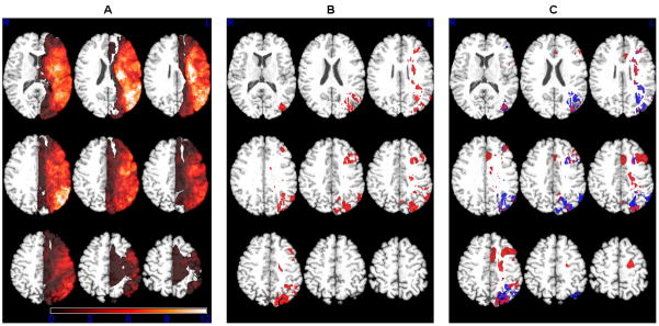
Oral reading is a complex skill involving the interaction of orthographic, phonological, and semantic processes. Functional imaging studies with nonimpaired adult readers have identified a widely distributed network of frontal, inferior parietal, posterior temporal, and occipital brain regions involved in the task. However, while functional imaging can identify cortical regions engaged in the process under examination, it cannot identify those brain regions essential for the task. The current study aimed to identify those neuroanatomical regions critical for successful oral reading by examining the relationship between word and nonword oral reading deficits and areas of tissue dysfunction in acute stroke. We evaluated 91 patients with left hemisphere ischemic stroke with a test of oral word and nonword reading, and magnetic resonance diffusion-weighted and perfusion-weighted imaging, within 24-48h of stroke onset. A voxel-wise statistical map showed that impairments in word and nonword reading were associated with a distributed network of brain regions, including the inferior and middle frontal gyri, the middle temporal gyrus, the supramarginal and angular gyri, and the middle occipital gyrus. In addition, lesions associated with word deficits were found to be distributed more frontally, while nonword deficits were associated with lesions distributed more posteriorly.
Copyright © 2010 Elsevier Inc. All rights reserved.
Figures
Similar articles
-
Voxel-based lesion analysis of brain regions underlying reading and writing.Neuropsychologia. 2018 Jul 1;115:51-59. doi: 10.1016/j.neuropsychologia.2018.03.021. Epub 2018 Mar 20. Neuropsychologia. 2018. PMID: 29572061 Free PMC article.
-
Neural systems for reading aloud: a multiparametric approach.Cereb Cortex. 2010 Aug;20(8):1799-815. doi: 10.1093/cercor/bhp245. Epub 2009 Nov 17. Cereb Cortex. 2010. PMID: 19920057 Free PMC article.
-
A neural network critical for spelling.Ann Neurol. 2009 Aug;66(2):249-53. doi: 10.1002/ana.21693. Ann Neurol. 2009. PMID: 19743449 Free PMC article.
-
Neural basis of single-word reading in Spanish-English bilinguals.Hum Brain Mapp. 2012 Jan;33(1):235-45. doi: 10.1002/hbm.21208. Epub 2011 Mar 9. Hum Brain Mapp. 2012. PMID: 21391265 Free PMC article.
-
Lesion analysis of the brain areas involved in language comprehension.Cognition. 2004 May-Jun;92(1-2):145-77. doi: 10.1016/j.cognition.2003.11.002. Cognition. 2004. PMID: 15037129 Review.
Cited by
-
Phonological and surface dyslexia in individuals with brain tumors: Performance pre-, intra-, immediately post-surgery and at follow-up.Hum Brain Mapp. 2020 Dec;41(17):5015-5031. doi: 10.1002/hbm.25176. Epub 2020 Aug 28. Hum Brain Mapp. 2020. PMID: 32857483 Free PMC article.
-
Brain regions that support accurate speech production after damage to Broca's area.Brain Commun. 2021 Oct 1;3(4):fcab230. doi: 10.1093/braincomms/fcab230. eCollection 2021. Brain Commun. 2021. PMID: 34671727 Free PMC article.
-
Contribution of writing to reading: Dissociation between cognitive and motor process in the left dorsal premotor cortex.Hum Brain Mapp. 2016 Apr;37(4):1531-43. doi: 10.1002/hbm.23118. Epub 2016 Jan 27. Hum Brain Mapp. 2016. PMID: 26813381 Free PMC article.
-
Targeted neurorehabilitation strategies in post-stroke aphasia.Restor Neurol Neurosci. 2023;41(3-4):129-191. doi: 10.3233/RNN-231344. Restor Neurol Neurosci. 2023. PMID: 37980575 Free PMC article. Review.
-
Four dimensions of naturalistic language production in aphasia after stroke.Brain. 2025 Jan 7;148(1):291-312. doi: 10.1093/brain/awae195. Brain. 2025. PMID: 38889230 Free PMC article.
References
-
- Banich MT. Executive function: The search for an integrated account. Current Directions in Psychological Science. 2009;18:89–94.
-
- Ben-Shachar M, Dougherty RF, Wandell BA. White matter pathways in reading. Current Opinion in Neurobiology. 2007;17:258–270. - PubMed
-
- Binder JR, McKiernan KA, Parsons ME, Westbury CF, Possing ET, Kaufman JN, Buchanan L. Neural correlates of lexical access during visual word recognition. Journal of Cognitive Neuroscience. 2003;15:372–393. - PubMed
-
- Brunner E, Munzel U. The nonparametric Behrens-Fisher problem: Asymptotic theory and a small-sample approximation. Biometrical Journal. 2000;42:17–25.
-
- Bub D. Alexia and related reading disorders. Neurologic Clinics of North America. 2003;21:549–568. - PubMed
Publication types
MeSH terms
Grants and funding
LinkOut - more resources
Full Text Sources
Medical
