. 2010 Dec 10;285(50):38944-50.
doi: 10.1074/jbc.M110.160325. Epub 2010 Oct 1.

Plk3 functions as an essential component of the hypoxia regulatory pathway by direct phosphorylation of HIF-1alpha

Affiliations

Plk3 functions as an essential component of the hypoxia regulatory pathway by direct phosphorylation of HIF-1alpha

Dazhong Xu et al. J Biol Chem. .

Abstract

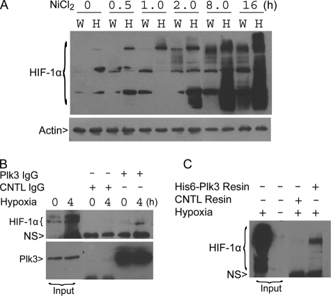
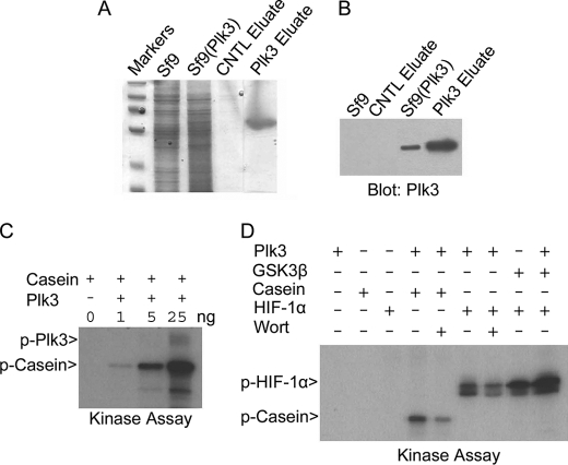
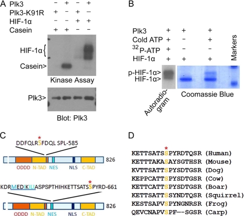
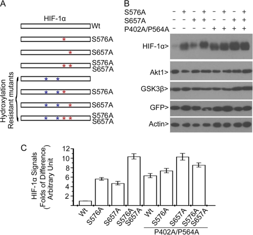
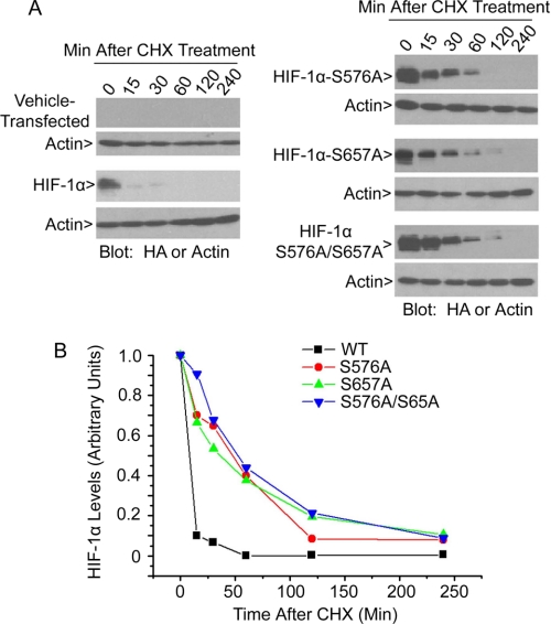
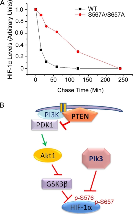
Polo-like kinase 3 (Plk3) plays an important role in the regulation of cell cycle progression and stress responses. Plk3 also has a tumor-suppressing activity as aging PLK3-null mice develop tumors in multiple organs. The growth of highly vascularized tumors in PLK3-null mice suggests a role for Plk3 in angiogenesis and cellular responses to hypoxia. By studying primary isogenic murine embryonic fibroblasts, we tested the hypothesis that Plk3 functions as a component in the hypoxia signaling pathway. PLK3(-/-) murine embryonic fibroblasts contained an enhanced level of HIF-1α under hypoxic conditions. Immunoprecipitation and pulldown analyses revealed that Plk3 physically interacted with HIF-1α under hypoxia. Purified recombinant Plk3, but not a kinase-defective mutant, phosphorylated HIF-1α in vitro, resulting in a major mobility shift. Mass spectrometry identified two unique serine residues that were phosphorylated by Plk3. Moreover, ectopic expression followed by cycloheximide or pulse-chase treatment demonstrated that phospho-mutants exhibited a much longer half-life than the wild-type counterpart, strongly suggesting that Plk3 directly regulates HIF-1α stability in vivo. Combined, our study identifies Plk3 as a new and essential player in the regulation of the hypoxia signaling pathway.


Figures

FIGURE 1.
FIGURE 1.
Plk3 functionally and physically interacts with HIF-1α. A, paired wild-type (W) and PLK3 homozygously disrupted (H) MEFs were treated with NiCl2, a hypoxia mimetic, for various times. Equal amounts of protein lysates were blotted for HIF-1α and β-actin. HIF-1α polyubiquitinated forms are indicated. B, A549 cells were treated with or without hypoxic stress for 4 h. An equal amount of cell lysates was immunoprecipitated with Plk3 IgG or with control IgG. After washing, the immunoprecipitates, along with the lysate inputs, were blotted for HIF-1α and Plk3. NS denotes a nonspecific band. C, recombinant His6-Plk3 was immobilized to Ni-NTA resin. His6-Plk3 resin, as well as control (CNTL) resin, was incubated with an equal amount of lysates from A549 cells exposed to the hypoxic stress. After thorough washing, proteins bound to either resin, along with lysate inputs, were blotted for HIF-1α. NS denotes a nonspecific band.
FIGURE 2.
FIGURE 2.
Plk3 phosphorylates HIF-1α in vitro. A, recombinant His6-Plk3 was expressed in Sf9 cells and purified using the Ni-NTA affinity approach as described under “Experimental Procedures.” Mock purification was carried out using Sf9 control cell lysates. Purified His6-Plk3 was confirmed by Coomassie Blue staining. B, Sf9 cell lysates infected with or without His6-Plk3 baculovirus, along with eluted proteins, were blotted with Plk3 antibody. C, different amounts of Plk3 were assayed for kinase activity toward casein in the kinase reaction supplemented with [γ-32P]ATP. A representative autoradiogram is shown. D, in vitro kinase assays were carried out in the presence of [γ-32P]ATP, as well as the various components, as indicated. Wort stands for wortmannin. A representative autoradiogram is shown.
FIGURE 3.
FIGURE 3.
Plk3 phosphorylates HIF-1α on Ser576 and Ser657. A, both Plk3 and Plk3-K91R were expressed in Sf9 cells and purified using NTA-Ni resin. Purified Plk3 and Plk3-K91R were assayed for their kinase activities toward HIF-1α in vitro in a kinase buffer containing [γ-32P]ATP. A representative autoradiogram is shown. B, kinase assays were carried out in the presence of various components as indicated. After the reaction, the samples were analyzed on SDS-PAGE followed by Coomassie Blue staining. A lane of “hot” sample (autoradiogram on the left) that ran on the same gel is also shown. C, HIF-1α sequence representation shows the relative positions of serine phosphorylation sites Ser576 (upper panel) and Ser657 (lower panel) to HIF-1α domains. ODDD, oxygen-dependent degradation domain; N-TAD, N-terminal transactivation domain; NES, nuclear export signal; NLS, nuclear localization signal; C-TAD, C-terminal transactivation domain. The nuclear export signal in the lower panel is highlighted by underlining. Ser641 and Ser643 are known to be phosphorylated by ERKs. D, amino acid alignment is shown of HIF-1α molecules from different species in the region that were targeted by Plk3. The conserved serine residues are highlighted.
FIGURE 4.
FIGURE 4.
Phosphorylation of HIF-1α by Plk3 leads to its destabilization. A, schematic representation of HIF-1α and its mutants (phospho- and/or hydroxylation mutants) used for transfection analyses is shown. B, HEK293 cells were co-transfected with various expression constructs as shown in C, and a GFP expression construct for normalization of transfection efficiency for 1 day is presented. Equal amounts of cell lysates were Western blotted for HIF-1α, Akt1, GSK3β, GFP, and β-actin. C, the signals shown in B were quantified by densitometry.
FIGURE 5.
FIGURE 5.
Plk3 phosphorylation-resistant mutants have longer half-lives than the wild-type HIF-1α. A, HEK293 cells were transfected with an HA-tagged HIF-1α expression construct or a construct expressing Plk3 phosphorylation-resistant mutant (HA-HIF-1α-S576A, HA-HIF-1α-S657A, or HA-HIF-1α-S576A/S657A) or vehicle for 1 day. Transfected cells were then treated with cycloheximide (CHX) for various times as indicated. Equal amounts of cell lysates from each treatment point were blotted for ectopically expressed HIF-1α protein using the anti-HA tag antibody. B, HIF-1α and its mutant protein signals shown in A were quantified by densitometry scanning, and the data were summarized from three independent experiments.
FIGURE 6.
FIGURE 6.
Plk3 functions as part of HIF-1α regulatory network. A, HEK293 cells transfected with HA-HIF-1α or with HA-HIF-1α-S576A/S657A for 48 h were pulsed with [35S]methionine/cysteine for 0.5 h followed by chase in an isotope-free medium for various periods of time. HEK293 cells were collected for lysate preparation. Equal amounts of lysates from various treatments were immunoprecipitated with the anti-HIF-1α antibody. The immunoprecipitates were fractionated on SDS-denaturing gels followed by autoradiography. HIF-1α-specific signals were qualified via densitometry, and the data are summarized. B, model is proposed illustrating how Plk3 regulates the hypoxia response network. Green arrows denote positive regulation; red bars denote negative regulation.

Similar articles

Cited by

References

    1. Barr F. A., Silljé H. H., Nigg E. A. (2004) Nat. Rev. Mol. Cell Biol. 5, 429–440 - PubMed
    1. Dai W. (2005) Oncogene 24, 214–216 - PubMed
    1. Zimmerman W. C., Erikson R. L. (2007) Cell Cycle 6, 1314–1318 - PubMed
    1. Zimmerman W. C., Erikson R. L. (2007) Proc. Natl. Acad. Sci. U.S.A. 104, 1847–1852 - PMC - PubMed
    1. Xie S., Wang Q., Wu H., Cogswell J., Lu L., Jhanwar-Uniyal M., Dai W. (2001) J. Biol. Chem. 276, 36194–36199 - PubMed

Publication types

Substances