. 2010 Dec 10;285(50):39401-8.
doi: 10.1074/jbc.M110.103705. Epub 2010 Oct 1.

Induction of interleukin-6 expression by bone morphogenetic protein-6 in macrophages requires both SMAD and p38 signaling pathways

Affiliations

Induction of interleukin-6 expression by bone morphogenetic protein-6 in macrophages requires both SMAD and p38 signaling pathways

Geun Taek Lee et al. J Biol Chem. .

Abstract

Unlike the prototype transforming growth factor-β (TGF-β), bone morphogenetic protein-6 (BMP-6) activates macrophages. Here, we report that BMP-6 induces the expression of IL-6 in macrophages. Using overexpression and knockdown experiments, we demonstrate that BMP receptor type II and activin-like kinase-2 are necessary for IL-6 induction by BMP-6. At the intracellular level, both Smad and p38 signaling pathways are required for the induction of IL-6. The cross-talk between the two pathways occurs at the level of transcription factor GATA4 and Smad 1/4. These results, taken together, demonstrate a novel BMP-6 signaling mechanism in which both the Smad and non-Smad pathways directly interact to activate the transcription of a target gene.


Figures

FIGURE 1.
FIGURE 1.
Induction of IL-6 expression by BMP-6 in macrophages. A, murine macrophage (Mφ) cell line RAW 264.7 and murine peritoneal macrophages were treated with increasing concentrations of BMP-6. RT-PCR (left panel) and immunoblot (right panel) analysis demonstrated that BMP-6 induced expression of IL-6 in macrophages in a concentration-dependent manner. B, RT-PCR (left panel) demonstrated that BMP-6 at 100 ng/ml induced expression of IL-6 mRNA within 1 h after treatment in RAW 264.7 and murine peritoneal macrophages. Immunoblot analysis (right panel) supported the results of RT-PCR. C, RAW 264.7 and murine peritoneal macrophages were treated with BMP-2, -4, -6, and -7 at 100 ng/ml, and IL-6 expression was measured by RT-PCR (top panel) and immunoblot analysis (bottom panel). Only BMP-6 induced IL-6 expression. D, RAW 264.7 and murine peritoneal macrophages were treated with cycloheximide (Cyclo) and actinomycin D (ActD) along with BMP-6 and IL-6 expression level was measured by RT-PCR. IL-6 mRNA induction by BMP-6 was blocked by actinomycin D but not by cycloheximide, suggesting that BMP-6 directly activates IL-6 promoter in macrophages. Con, control.
FIGURE 2.
FIGURE 2.
BMP-6 receptors and IL-6 induction in macrophages. A, plasmid IL-6-Luc containing IL-6 promoter and luciferase reporter was transfected into RAW 264.7 cells and treated with 100 ng/ml BMP-6. Twenty-four hours after adding BMP-6, IL-6 promoter activity increased more than 3-fold. B, constitutively active ALK-2 and -3 were co-transfected with IL-6-Luc reporter. Although both ALK-2 and -3 induced IL-6 promoter activity to a statistically significant level, ALK-2 increased IL-6 promoter activity more than 3-fold. C, lentiviruses containing shRNA sequences targeting ALK-2 and -3 were infected into RAW 264.7 cells. Statistically significant knockdown of target gene expression was confirmed previously and published (13). When transfected with IL-6-Luc and treated with BMP-6, only the knockdown of ALK-2 blocked the induction of luciferase activity. D, macrophages express all three known BMP type II receptors (BMP-RII, Act-RIIA, and Act-RIIB). Thus, each of the three type II BMP receptors was co-transfected with IL-6-Luc into RAW 264.7 cells and treated with BMP-6. Only in cells transfected with BMP-RII was a significant increase in IL-6 promoter activity observed. Interestingly, overexpression of Act-RIIB suppressed IL-6 promoter activity consistently. E, lentiviruses containing shRNA sequences targeting each of the three type II BMP receptors were infected into RAW 264.7 cells. We have previously confirmed the knockdown of the target gene expression (13). Among the three type II BMP receptors, knockdown of BMP-RII reversed the induction of IL-6 promoter. *, statistically significant.
FIGURE 3.
FIGURE 3.
Smads and IL-6 induction in macrophages. A, BMPs signal through the R-Smads 1, 5, and 8. Confocal microscopy in peritoneal macrophages demonstrated that BMP-6 induced increased levels as well as nuclear translocation of phospho-Smad 1/5. B, dominant negative Smad 4 (Smad4DN) was transfected into RAW 264.7 cells and treated with increasing concentrations of BMP-6. Effect on IL-6 expression was measured using RT-PCR. Transfection process itself modestly increased the baseline expression level of IL-6. Compared with the control, IL-6 induction was suppressed in cells expressing Smad4DN, suggesting that the Smad-dependent pathway is necessary for IL-6 induction in macrophages. C, RAW 264.7 cells were co-transfected with each of the three R-Smads (Smad 1, 5, and 8) and the Co-Smad (Smad 4) along with IL-6-Luc. When treated with BMP-6, cells expressing Smad 1/4 demonstrated the highest level of induction of IL-6 promoter activity. Interestingly, transfection with Smad 8/4 resulted in suppression of IL-6 promoter activity. D, lentiviruses containing shRNA sequences against Smad 1, 5, and 8 were infected into RAW 264.7 cells. Statistically significant knockdown of target gene expression was confirmed using RT-PCR (supplemental Fig. 2). When transfected with IL-6-Luc and treated with BMP-6, knockdown of Smad 1 blocked the induction of luciferase activity whereas that of Smad 8 increased luciferase activity. E, to determine the BMP-6-response element in the IL-6 promoter region, serial deletion constructs of IL-6 promoter were established. Subsequently, the shortened IL-6 promoters were co-transfected with IL-6-Luc into RAW 264.7 cells. In IL-6 promoter, BMP-6-response element was located between −50 and −150 bp 5′ to the transcription initiation site. The results shown are induced luciferase activity by BMP-6. F, ChIP was carried out to determine the presence of interaction between Smads and IL-6 promoter. The −50 to −150 region was amplified in samples immunoprecipitated with Smad 1 and 4 antibodies. As a negative control, the −150 to −300 bp region was targeted. *, statistically significant.
FIGURE 4.
FIGURE 4.
Non-Smad p38 pathway and IL-6 induction in macrophages. A, immunoblot analysis was carried out to determine the effect of BMP-6 on activation of p38. BMP-6 induced the phosphorylation of p38 within 5–15 min in RAW 264.7 and murine peritoneal macrophages. p-p38 = phosphorylated p38; t-p38, total p38. B, immunofluorescence microscopy was used to localize p38 following stimulating with BMP-6 in RAW 264.7. The cytosolic location of p38 did not change with BMP-6 treatment. C, to determine the role of p38 activation on IL-6 induction by BMP-6 in macrophages, RAW 264.7 cells were treated with 10 μm SB 203580, a p38 inhibitor. RT-PCR demonstrated that IL-6 induction was blocked when p38 was inhibited. D, dominant negative p38 (p38DN) was transfected into RAW 264.7 cells, and the effect on IL-6 was measured using RT-PCR. As with cells treated with SB 203580, expression of p38DN blocked the induction of IL-6. E, a classic activator of IL-6 expression is NF-κB. Thus, RAW 264.7 was treated with BAY11-7082 to inhibit the NF-κB pathway. When simultaneously treated with BMP-6, RT-PCR showed no significant effect on IL-6 induction. These results demonstrate that IL-6 induction by BMP-6 does not require the NF-κB pathway. F, to determine the effect of p38 activation on Smad pathway, immunofluorescence microscopy was used. The results revealed that the inhibition of the p38 pathway via SB 203580 had no impact on nuclear translocation of phosphorylated Smad 1/5 (pSmad 1/5).
FIGURE 5.
FIGURE 5.
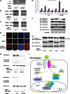
GATA 4 and IL-6 expression in macrophages. A, RAW 264.7 was transfected with GATA4 siRNA (siGATA4), and the effect on IL-6 expression was measured using RT-PCR. When treated with BMP-6, induction of IL-6 was no longer observed when GATA4 was knocked down. B, ChIP was carried out to determine the interaction between GATA4 and the IL-6 promoter. GATA3 was used as a control. Following treatment with BMP-6, the −50 to −150 bp region was amplified in samples immunoprecipitated with GATA4 but not GATA3 antibody. C, when RAW 264.7 cells were treated with SB 203580 and BMP-6, ChIP assay using GATA4 antibody no longer amplified the −50 to −150 bp region. This observation demonstrates that GATA4 signals downstream of p38. D, confocal immunofluorescence microscopy was carried out using antibodies against Smad 1 and GATA 4. BMP-6 treatment induced nuclear translocation of Smad 1 and GATA4 simultaneously. E, RAW 264.7 was transfected with indicated combination of Smad 1, 4, 5, and 8 and GATA4 along with IL-6-Luc. BMP-6 induced a statistically significant level of IL-6 promoter activity when GATA4 was co-transfected with Smad 1/4. However, transfection of GATA4 alone did not induce IL-6 promoter activity (control bar in GATA4 group). F, myc-tagged Smads 1, 4, 5, and 8, along with FLAG-tagged GATA4 were expressed in RAW 264.7. Immunoprecipitation using anti-FLAG antibody followed by immunoblot analysis against myc epitope was performed. The results demonstrated that Smads 1, 4, and 8 but not 5 interacted with GATA4. G, myc-tagged Smads 1, 4, 5, and 8 were expressed in RAW 264.7. Immunoprecipitation using anti-myc antibody followed by immunoblot against endogenous GATA4 was performed. Following treatment with BMP-6, increased levels of GATA4 protein were immunoprecipitated out from cells transfected with Smad 1 or 4. H, interaction between endogenous Smad 1 and GATA4 was investigated using the shRNA approach. When Smad 1 was knocked down and GATA4 was immunoprecipitated, immunoblot for Smad 1 revealed no protein band. Conversely, when GATA4 was knocked down and Smad 1 was immunoprecipitated, immunoblotting for GATA4 demonstrated no protein band. I, proposed mechanism of IL-6 induction by BMP-6 in macrophages. Induction of IL-6 expression by BMP-6 in macrophages requires both Smad and non-Smad pathways. BMP-RII along with ALK2 simultaneously activates Smad 1/4 and p38. Subsequently, p38 activates GATA4. Finally, GATA4 and Smad 1/4 translocate to the nucleus and bind to the BMP-6-response element within IL-6 promoter and induce IL-6 expression. *, statistically significant.

Similar articles

Cited by

References

    1. Wozney J. M., Rosen V., Celeste A. J., Mitsock L. M., Whitters M. J., Kriz R. W., Hewick R. M., Wang E. A. (1988) Science 242, 1528–1534 - PubMed
    1. Zhao G. Q. (2003) Genesis 35, 43–56 - PubMed
    1. Miyazono K., Maeda S., Imamura T. (2005) Cytokine Growth Factor Rev. 16, 251–263 - PubMed
    1. Liu F., Ventura F., Doody J., Massagué J. (1995) Mol. Cell. Biol. 15, 3479–3486 - PMC - PubMed
    1. Kretzschmar M., Massagué J. (1998) Curr. Opin. Genet. Dev. 8, 103–111 - PubMed

Publication types

LinkOut - more resources