. 2010 Nov;177(5):2387-98.
doi: 10.2353/ajpath.2010.100475. Epub 2010 Oct 1.

Sera from preeclampsia patients elicit symptoms of human disease in mice and provide a basis for an in vitro predictive assay

Affiliations

Sera from preeclampsia patients elicit symptoms of human disease in mice and provide a basis for an in vitro predictive assay

Satyan Kalkunte et al. Am J Pathol. 2010 Nov.

Abstract

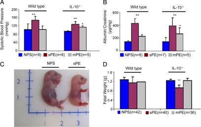
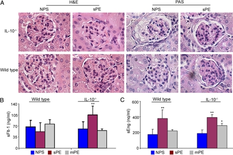
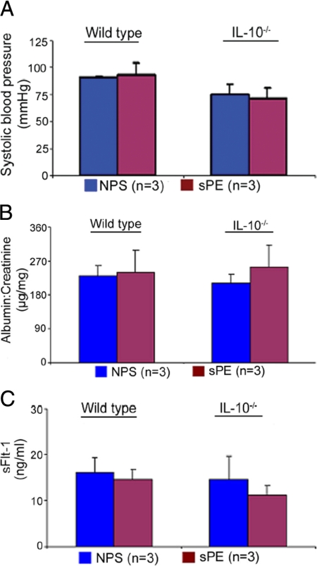
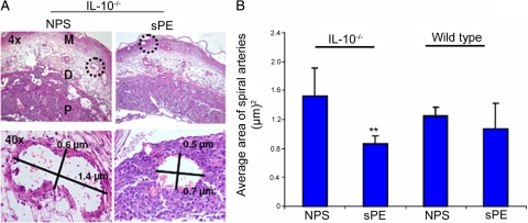
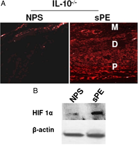
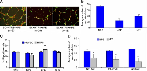
Early diagnosis and treatment of preeclampsia would significantly reduce maternal and fetal morbidity and mortality. However, its etiology and prediction have remained elusive. Based on the hypothesis that sera from patients with preeclampsia could function as a "blueprint" of causative factors, we describe a serum-based pregnancy-specific mouse model that closely mirrors the human condition as well as an in vitro predictive assay. We show that a single administration of human preeclampsia serum in pregnant IL-10-/- mice induced the full spectrum of preeclampsia-like symptoms, caused hypoxic injury in uteroplacental tissues, and elevated soluble fms-like tyrosine kinase 1 and soluble endoglin, markers thought to be related to the disease. The same serum sample(s) induced a partial preeclampsia phenotype in wild-type mice. Importantly, preeclampsia serum disrupted cross talk between trophoblasts and endothelial cells in an in vitro model of endovascular activity. Disruption of endovascular activity could be documented in serum samples as early as 12 to 14 weeks of gestation from patients who subsequently developed preeclampsia. These results indicate that preeclampsia patient sera can be used to understand the pregnancy-specific disease pathology in mice and can predict the disorder.


Figures

Figure 1
Figure 1
Preeclampsia serum induces symptoms of hypertension, proteinuria, and IUGR in pregnant IL-10−/− mice. Pregnant wild-type or IL-10−/− mice were injected with normal pregnancy serum (NPS), severe preeclampsia serum (sPE), or mild preeclampsia serum (mPE) on gd 10. As depicted in the figure, multiple serum samples (parentheses) were used in multiple mating experiments. A: Systolic blood pressures of pregnant mice in response to different treatments are shown. Treatment with sPE (magenta bar) induces significant hypertension in both wild-type and IL-10−/− mice when compared with treatment with NPS (blue bar). Treatment with mPE (gray bar) induces moderate hypertension only in IL-10−/− mice compared with the NPS group. B: Proteinuria values from urine samples collected over 24-hour periods in response to different treatments are expressed as albumin to creatinine (microgram per milligram) ratios. Treatment with sPE (magenta bar) induces significant proteinuria in both wild-type and IL-10−/− mice compared with treatment with NPS (blue bar). Treatment with mPE (gray bar) induces moderate proteinura in IL-10−/− mice without significant effect in wild-type counterparts. C: A representative photograph of a gd 17 fetus shows sPE-induced growth restriction compared with that with NPS treatment. D: Average weights of a number of fetuses (n) from wild-type or IL-10−/− mice are represented for different treatment groups. Treatment with sPE (magenta bar), but not mPE (gray bar) or NPS (blue bar), induces IUGR in IL-10−/− mice with no significant effect in wild-type counterparts. All values represent the mean ± SD of at least seven animals per group. The numbers in parentheses indicate the number of serum samples tested in the study. *P < 0.01; **P < 0.05 for PE or normal pregnancy serum groups.
Figure 2
Figure 2
Preeclampsia serum induces glomerular endotheliosis and elevated sFlt-1 and sEng production. A: Histopathological analyses of renal tissue from representative NPS- or sPE-treated pregnant IL-10−/− (upper panel) and wild-type mice (lower panel) are shown (original magnification, ×100). H&E stain shows capillary occlusion in the sPE-treated IL-10−/− and wild-type animals with enlarged glomeruli and swollen endothelial cells compared with NPS-treated control mice. PAS-based staining of the sPE-treated mice shows inflammation of capillary endothelial cells (endotheliosis) in both IL-10−/− and wild-type animals. These pathological changes are absent in the NPS-treated mice. Similar results were observed with multiple serum samples. A representative image from the staining of at least three animals per group is shown. B: Circulating gd 17 mouse serum levels of sFlt-1 in response to a single-dose treatment with NPS (blue bar), sPE (magenta bar), and mPE (gray bar) are shown. Treatment with sPE induces excess production of sFlt-1 in IL-10−/− mice compared with NPS treatment without influencing wild-type counterparts. Treatment with mPE serum did not induce the production of sFlt-1 in either wild-type or IL-10−/− mice. C: Circulating gd 17 mouse serum levels of sEng in response to treatment with NPS, sPE, or mPE are shown in the graph. Treatment with sPE induces excess production of sEng both in IL-10−/− mice as well as wild-type animals when compared with NPS treatment. Treatment with mPE induced moderate increase in the levels of sEng only in IL-10−/− mice. All values are expressed as the mean ± SD obtained from at least seven animals per treatment group. *P < 0.01 or **P < 0.05 represent significance over control NPS-treated groups.
Figure 3
Figure 3
Severe preeclampsia serum does not induce disease-like symptoms in nonpregnant mice. Nonpregnant wild-type or IL-10−/− female mice were injected with NPS or sPE samples. Seven days after a single administration as described for pregnant mice, blood pressure was monitored, and urine and serum samples were analyzed. A: No changes were observed in the readings of systolic blood pressure in nonpregnant mice irrespective of treatment with NPS (blue bar) or sPE (magenta bar). B: Proteinuria values from urine samples collected over a 24-hour period in response to different treatments are shown. Treatment with sPE did not induce significant proteinuria in either wild-type or IL-10−/− mice compared with NPS treatment. C: Circulating mouse serum levels of soluble Flt-1(sFlt-1) in response to a single-dose treatment of NPS or sPE are shown. No significant differences were observed between NPS (blue bar) and sPE (magenta bar). All of the values were obtained from at least five animals per treatment group and are expressed as the mean ± SD. The numbers in parentheses indicate the number of different serum samples tested in the study.
Figure 4
Figure 4
Preeclampsia serum blocks the spiral artery remodeling in pregnant IL-10−/− mice. Pregnant wild-type and IL-10−/− were injected with NPS, or sPE on gd 10, and uteroplacental units were analyzed on gd 13 as described in Materials and Methods. A: H&E staining of a uteroplacental section from one representative NPS- or sPE-treated pregnant IL-10−/− mouse is shown (upper panel; original magnification, ×4), and the representative morphometry of a spiral artery indicated by a dotted circle is shown at higher magnification (×40, lower panel). The lengths and widths of spiral arteries expressed in micrometers (μm) were measured and stamped by using SPOT Advanced software. Treatment with sPE resulted in poor remodeling of spiral arteries as indicated by their reduced size compared with NPS-treated control animals. B represents the average area of the spiral arteries of at least six spiral arteries per placental unit from three independent animals per treatment group. **P < 0.05 compared with the NPS-treated group. M, mesometrium; D, decidua basalis; P, placenta.
Figure 5
Figure 5
Preeclampsia serum induces placental hypoxia in IL-10−/− mice. Pregnant wild-type and IL-10−/− mice were injected with NPS or sPE on gd 10 (n = 3 each), and uteroplacental units were analyzed on gd 13. Animals were injected with EF5 as described in Materials and Methods. A: Immunofluorescence of a representative uteroplacental section stained with an EF5 specific antibody (original magnification, ×10). Treatment with sPE significantly induced hypoxia in IL-10−/− mice compared with NPS-treated mice. The treatment with sPE did not induce significant hypoxic injury in wild-type uteroplacental tissue. Note that EF5 staining was more prominent in the placental region (P) and that hypoxic injury was also visible in the decidua (D) and around the spiral arteries in the mesometrial region (M). B: Expression of HIF 1α in response to treatment with NPS and sPE in IL-10−/− placenta harvested on gd 12 is shown in a representative Western blot. β-actin is used as a loading control. Treatment with sPE significantly induced HIF 1α compared with NPS treatment and confirms the hypoxic effects seen with EF5 staining. These data represent at least three experiments.
Figure 6
Figure 6
Preeclampsia serum disrupts endovascular interaction between trophoblasts and endothelial cells in vitro. A: Human umbilical vein endothelial cells (EC; labeled red) and first-trimester human trophoblasts (HTR8; labeled green) were cultured overnight on Matrigel in the presence of gestational age-matched NPS, sPE, or mPE serum. Capillary tube formation was recorded as described in Materials and Methods. Representative figures of EC-directed tube formation by HTR8 cells (original magnification, ×4) are shown. NPS significantly supported tube formation, whereas sPE and mPE serum disrupted this interaction, albeit in a mild or severe pathology-dependent manner. B shows the quantification of dual cell endovascular tube formation in response to different serum samples. The average number of tubes/vacuoles formed was quantified by counting the number of tube-like structures formed by connected capillary bridge in four different fields (original magnification, ×4) as described in Materials and Methods and represents multiple experiments. C: Cytotoxic potential of NPS and sPE on EC and HTR8 by propidium iodide (PI) staining was analyzed by FACS and represented here as % PI positive cells. Treatment with sPE or NPS did not induce cytotoxicity in either EC or HTR8. D: Longitudinal studies involving pregnancy serum samples from weeks 12 to 14 (n = 18), 24 to 27 (n = 5), or 32 to 36 (n = 22) weeks of gestation were tested for supporting tube formation. These samples were from women who were later diagnosed with preeclampsia. Data are represented as a comparative analysis between these samples and those from gestational age-matched normal pregnancies. The average number of tubes/vacuoles formed was quantified and plotted. As seen in D, unlike NPS, the tube disrupting activity in preeclampsia serum could be traced back to 12 to 14 weeks of pregnancy. All of the values are expressed as the mean ± SD. *P < 0.01 or **P < 0.05 compared with the respective control NPS-treated group.

Similar articles

Cited by

References

    1. Sibai B, Dekker G, Kupferminc M. Pre-eclampsia. Lancet. 2005;365:785–799. - PubMed
    1. Redman CW, Sargent IL. Latest advances in understanding preeclampsia. Science. 2005;308:1592–1594. - PubMed
    1. Ilekis JV, Reddy UM, Roberts JM. Preeclampsia— a pressing problem: an executive summary of a National Institute of Child Health and Human Development workshop. J Reprod Sci. 2007;14:508–523. - PubMed
    1. Noris M, Perico N, Remuzzi G. Mechanisms of disease: pre-eclampsia. Nat Clin Pract Nephrol. 2005;1(2):98–114. - PubMed
    1. Redman CWG, Sargent IL. Placental debris, oxidative stress and preeclampsia. Placenta. 2000;21:597–602. - PubMed

Publication types

MeSH terms

Substances