. 2011 Jan;25(1):358-69.
doi: 10.1096/fj.10-171579. Epub 2010 Oct 1.

Macrophages recruited via CCR2 produce insulin-like growth factor-1 to repair acute skeletal muscle injury

Affiliations

Macrophages recruited via CCR2 produce insulin-like growth factor-1 to repair acute skeletal muscle injury

Haiyan Lu et al. FASEB J. 2011 Jan.

Abstract

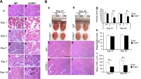
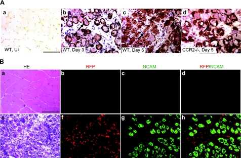
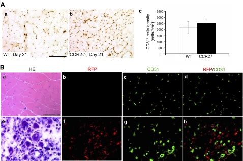
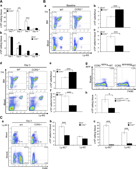
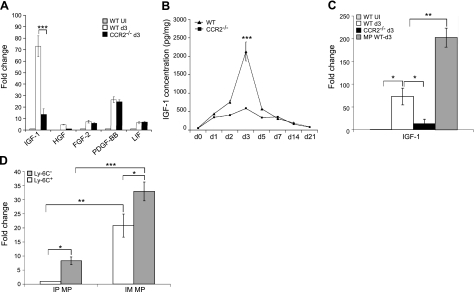
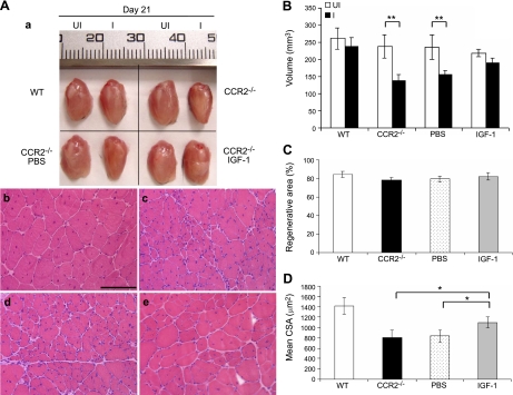
CC chemokine receptor 2 (CCR2) is essential to acute skeletal muscle injury repair. We studied the subpopulation of inflammatory cells recruited via CCR2 signaling and their cellular functions with respect to muscle regeneration. Mobilization of monocytes/macrophages (MOs/MPs), but not lymphocytes or neutrophils, was impaired from bone marrow to blood and from blood to injured muscle in Ccr2(-/-) mice. While the Ly-6C(+) but not the Ly-6C(-) subset of MOs/MPs was significantly reduced in blood, both subsets were drastically reduced in injured muscle of Ccr2(-/-) mice. Expression of insulin-like growth factor-1 (IGF-I) was markedly up-regulated in injured muscle of wild-type but not Ccr2(-/-) mice. IGF-I was strongly expressed by macrophages within injured muscle, more prominently by the Ly-6C(-) subset. A single injection of IGF-I, but not PBS, into injured muscle to replace IGF-I remarkably improved muscle regeneration in Ccr2(-/-) mice. CCR2 was not detected in myogenic cells or capillary endothelial cells in injured muscle to suggest its direct involvement in muscle regeneration or angiogenesis. We conclude that CCR2 is essential to acute skeletal muscle injury repair primarily by recruiting Ly-6C(+) MOs/MPs. Within injured muscle, these cells conduct phagocytosis, contribute to accumulation of intramuscular Ly-6C(-) macrophages, and produce a high level of IGF-I to promote muscle regeneration.


Figures

Figure 1.
Figure 1.
Ccr2−/− mice showed markedly reduced muscle inflammation, delayed phagocytosis, and impaired muscle regeneration after acute injury induced by BaCl2. A) Hematoxylin-eosin staining showed myofiber necrosis and tissue edema in injured muscle at d 1 of both wild-type (c) and Ccr2−/− mice (d). While robust inflammatory infiltrates were seen at d 3 in injured muscle of wild-type mice (e), very few inflammatory cells were seen in Ccr2−/− mice (f). Necrotic fibers were gradually replaced by mononucleated and multinucleated myoblasts and myotubes at d 5 (g) and 7 (i) in wild-type mice; they remained in Ccr2−/− mice with scattered endomysial inflammation (h, j). While the injured areas were filled with central-nucleated regenerated myofibers with no necrotic fibers or inflammatory cells seen in wild-type mice at d 14 (k), scattered necrotic fibers (arrows) and inflammatory cells were still present in Ccr2−/− mice along with smaller regenerated fibers (l). B) At d 21 and 42, the injured muscle (a, b) and regenerated muscle fibers (c-f) were smaller in Ccr2−/− mice (a, b, e, f) than in wild-type mice (a–d). Quantitative analyses showed that the volume of injured muscle (g) and the mean CSA of regenerated fibers (i) were significantly reduced in Ccr2−/− mice as compared with wild-type controls. There was no difference in the percentage of regenerative area (h). n = 5–7 mice/group/time point. WT, wild-type; UI, uninjured; I, injured. Scale bars = 50 μm. **P < 0.01.
Figure 2.
Figure 2.
CCR2 was not expressed on myogenic cells in injured muscle.. A) Immunostaining of NCAM showed no positive staining in normal muscle (a). In injured muscle of wild-type mice at d 3, satellite cells were activated expressing NCAM (b, arrow). At d 5, many NCAM+ myoblasts and multinucleated myotubes were also seen (c, arrow). In Ccr2−/− mice at d 5, only activated satellite cells and small myoblasts were seen surrounding necrotic fibers (d, arrow). B) NCAM/RFP double immunostaining of injured muscle of Ccr2RFP/+ mice at d 5 showed RFP+ inflammatory cells (f) and NCAM membrane staining of myogenic cells (g). No RFP+/NCAM+ cells were detected (h) to indicate expression of CCR2 on myogenic cells. RFP or NCAM was not detected in uninjured muscle of Ccr2RPP/+ mice (b–d). Adjacent sections of uninjured muscle (a) and injured muscle at d 5 (e) were stained with hematoxylin-eosin (HE). n = 3 mice/group. Scale bars = 50 μm.
Figure 3.
Figure 3.
CCR2 was not expressed on capillary endothelial cells in injured muscle.. A) CD31+ cell densities were not significantly different in wild-type (a, c) and Ccr2−/− mice (b, c) at d 21. B) RFP/CD31 double immunostaining of uninjured (b–d) and injured (f–h) muscle of Ccr2RFP/+ mice showed that no RFP+/CD31+ cells were detected in uninjured (d) or injured muscle (h) at d 5 to indicate expression of CCR2 on capillary endothelial cells. Adjacent sections of uninjured muscle (a) and injured muscle at d 5 (e) were stained with hematoxylin-eosin (HE). Bar = 50 μm; n = 3 mice/group.
Figure 4.
Figure 4.
CCR2 deficiency blocked MO/MP recruitment into injured muscle.. A) Flow cytometry showed a markedly increased number of intramuscular macrophages in wild-type mice at d 3 and 7, with the peak at d 3 (a). The number of intramuscular macrophages in Ccr2−/− mice was also increased after injury (a) but was significantly reduced at d 3 and 7 as compared with wild-type mice. The number of neutrophils in Ccr2−/− mice was increased at d 3 as compared with wild-type mice (b). B) Flow cytometry at baseline showed that the percentage of 7/4+Ly-6G MOs/MPs (a) was increased in bone marrow but reduced in blood in Ccr2−/− mice as compared with wild-type controls. At d 3 postinjury, the percentage of 7/4+Ly-6G MOs/MPs (d) was also increased in bone marrow and reduced in blood in Ccr2−/− mice. The numbers of 7/4+Ly-6G MOs/MPs per femur were significantly higher in bone marrow (BM; b, e) but lower in blood (c, f) at baseline (b, c) and d 3 (e, f) in Ccr2−/− mice than those in wild-type mice. Bone marrow monocyte transfer showed that the number of Ccr2RFP/+ monocytes recruited into wild-type injured muscle at d 3 was significantly higher than that of Ccr2RFP/RFP monocytes (lacking CCR2) (g, h). C) Flow cytometry at d 3 showed that the numbers of F4/80+ MOs/MPs were reduced in blood and in injured muscle of Ccr2−/− mice (a), among which the Ly-6C+ but not the Ly-6C subset was significantly reduced in blood (b). However, both Ly-6C+ and Ly-6C subsets were significantly reduced in injured muscle of Ccr2−/− mice (c). n = 5–7 mice/group/experiment. *P < 0.05; **P < 0.01; ***P < 0.001.
Figure 5.
Figure 5.
Macrophages (MPs) expressed a high level of IGF-I in injured muscle. A) Quantitative RT-PCR showed that the IGF-I mRNA expression was significantly higher in injured muscle of wild-type mice than in those of Ccr2−/− mice at d 3. B) ELISA analysis showed that the level of IGF-I was increased in injured muscle of both wild-type and Ccr2−/− mice, with the peak at d 3, at which point the IGF-I level was remarkably higher in wild-type mice than in Ccr2−/− mice. C) Quantitative RT-PCR showed that the MPs from injured muscle of wild-type mice at d 3 expressed a remarkably higher level of IGF-I mRNA than the injured muscle itself from wild-type or Ccr2−/− mice. D) Quantitative RT-PCR showed that both Ly-6C+ and Ly-6C intramuscular MPs (IM MP) expressed IGF-I mRNA, the level being significantly higher in the Ly-6C subset than in the Ly-6C+ subset. Although the intraperitoneal MPs (IP MP) also expressed IGF-I mRNA, the level was much lower than that in the IM MPs. n = 5–7 mice/group/experiment. Fold change refers to the comparison to wild-type uninjured (UI) muscle (A, C) and to Ly-6C+ IP MPs (D). *P < 0.05; ** P < 0.01; ***P < 0.001.
Figure 6.
Figure 6.
Local IGF-I treatment improved muscle regeneration in Ccr2−/− mice. At d 21, while the injured quadriceps muscle of wild-type mice regenerated to reach the size of contralateral uninjured controls, the size (volume) of injured quadriceps in Ccr2−/− mice was significantly smaller than the contralateral uninjured controls (Aa, B). Regenerated muscle fibers were also smaller in Ccr2−/− mice (Ac) than in wild-type mice (Ab). Intramuscular injection of IGF-I (Aa, e; B; D), but not PBS (Aa, d; B; D), improved muscle regeneration with increased muscle volume and muscle fiber size. There was no difference in the percentage of regenerative area in these muscles (C). n = 5 mice/group/experiment. Scale bar = 50 μm. *P < 0.05; **P < 0.01.

Similar articles

Cited by

References

    1. Contreras-Shannon V., Ochoa O., Reyes-Reyna S. M., Sun D., Michalek J. E., Kuziel W. A., McManus L. M., Shireman P. K. (2007) Fat accumulation with altered inflammation and regeneration in skeletal muscle of CCR2−/− mice following ischemic injury. Am. J. Physiol. Cell Physiol. 292, C953–C967 - PubMed
    1. Arnold L., Henry A., Poron F., Baba-Amer Y., van Rooijen N., Plonquet A., Gherardi R. K., Chazaud B. (2007) Inflammatory monocytes recruited after skeletal muscle injury switch into anti-inflammatory macrophages to support myogenesis. J. Exp. Med. 204, 1057–1069 - PMC - PubMed
    1. Sun D., Martinez C. O., Ochoa O., Ruiz-Willhite L., Bonilla J. R., Centonze V. E., Waite L. L., Michalek J. E., McManus L. M., Shireman P. K. (2009) Bone marrow-derived cell regulation of skeletal muscle regeneration. FASEB J. 23, 382–395 - PMC - PubMed
    1. Tidball J. G. (2005) Inflammatory processes in muscle injury and repair. Am. J. Physiol. Regul. Integr. Comp. Physiol. 288, R345–353 - PubMed
    1. Bischoff R., Heintz C. (1994) Enhancement of skeletal muscle regeneration. Dev. Dyn. 201, 41–54 - PubMed

Publication types

MeSH terms

LinkOut - more resources